• 文献检索
  • 文档翻译
  • 深度研究
  • 学术资讯
  • Suppr Zotero 插件Zotero 插件
  • 邀请有礼
  • 套餐&价格
  • 历史记录
应用&插件
Suppr Zotero 插件Zotero 插件浏览器插件Mac 客户端Windows 客户端微信小程序
定价
高级版会员购买积分包购买API积分包
服务
文献检索文档翻译深度研究API 文档MCP 服务
关于我们
关于 Suppr公司介绍联系我们用户协议隐私条款
关注我们

Suppr 超能文献

核心技术专利:CN118964589B侵权必究
粤ICP备2023148730 号-1Suppr @ 2026

文献检索

告别复杂PubMed语法,用中文像聊天一样搜索,搜遍4000万医学文献。AI智能推荐,让科研检索更轻松。

立即免费搜索

文件翻译

保留排版,准确专业,支持PDF/Word/PPT等文件格式,支持 12+语言互译。

免费翻译文档

深度研究

AI帮你快速写综述,25分钟生成高质量综述,智能提取关键信息,辅助科研写作。

立即免费体验

无需使用造影剂的灌注毛细血管高分辨率宽视野成像。

High-resolution wide-field imaging of perfused capillaries without the use of contrast agent.

作者信息

Nelson Darin A, Burgansky-Eliash Zvia, Barash Hila, Loewenstein Anat, Barak Adiel, Bartov Elisha, Rock Tali, Grinvald Amiram

机构信息

Optical Imaging Ltd, Rehovot, Israel;

出版信息

Clin Ophthalmol. 2011;5:1095-106. doi: 10.2147/OPTH.S20103. Epub 2011 Aug 9.

DOI:10.2147/OPTH.S20103
PMID:21887088
原文链接:https://pmc.ncbi.nlm.nih.gov/articles/PMC3162286/
Abstract

PURPOSE

Assessment of capillary abnormalities facilitates early diagnosis, treatment, and follow-up of common retinal pathologies. Injected contrast agents like fluorescein are widely used to image retinal capillaries, but this highly effective procedure has a few disadvantages, such as untoward side effects, inconvenience of injection, and brevity of the time window for clear visualization. The retinal function imager (RFI) is a tool for monitoring retinal functions, such as blood velocity and oximetry, based on intrinsic signals. Here we describe the clinical use of hemoglobin in red blood cells (RBCs) as an intrinsic motion-contrast agent in the generation of detailed noninvasive capillary-perfusion maps (nCPMs).

PATIENTS AND METHODS

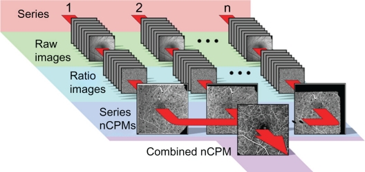
Multiple series of nCPM images were acquired from 130 patients with diabetic retinopathy, vein occlusion, central serous retinopathy, age-related macular degeneration, or metabolic syndrome, as well as from 37 healthy subjects. After registration, pixel value distribution parameters were analyzed to locate RBC motion.

RESULTS

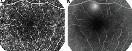
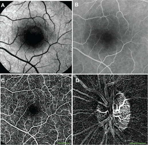
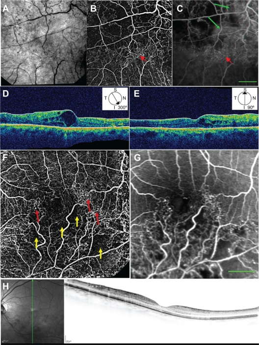
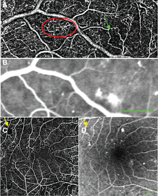
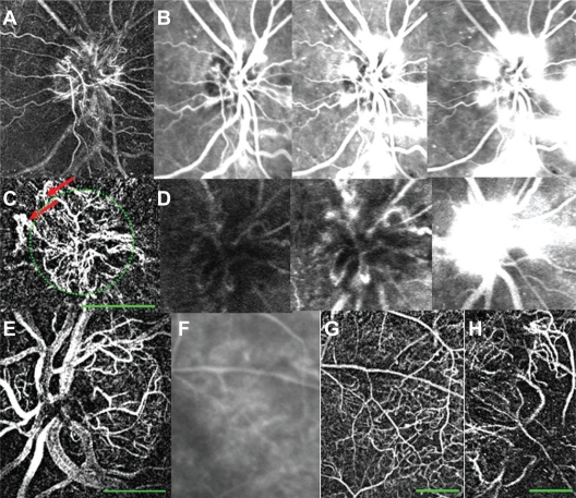
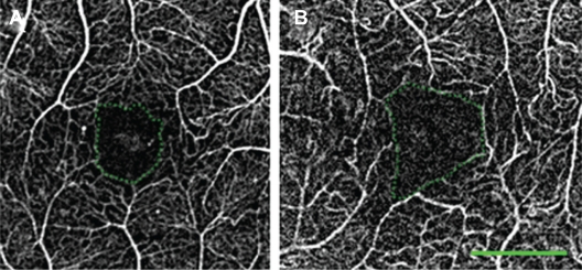
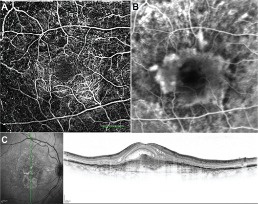
The RFI yielded nCPMs demonstrating microvascular morphology including capillaries in exquisite detail. Maps from the same subject were highly reproducible in repeated measurements, in as much detail and often better than that revealed by the very best fluorescein angiography. In patients, neovascularization and capillary nonperfusion areas were clearly observed. Foveal avascular zones (FAZ) were sharply delineated and were larger in patients with diabetic retinopathy than in controls (FAZ diameter: 641.5 ± 82.3 versus 463.7 ± 105 μm; P < 0.001). Also visible were abnormal vascular patterns, such as shunts and vascular loops.

CONCLUSION

Optical imaging of retinal capillaries in human patients based on motion contrast is noninvasive, comfortable, safe, and can be repeated as often as required for early diagnosis, treatment guidance, and follow up of retinal disease progression.

摘要

目的

评估毛细血管异常有助于常见视网膜病变的早期诊断、治疗及随访。像荧光素这样的注射用造影剂被广泛用于视网膜毛细血管成像,但这种高效的方法存在一些缺点,如不良副作用、注射不便以及清晰可视化的时间窗较短。视网膜功能成像仪(RFI)是一种基于内在信号监测视网膜功能(如血流速度和血氧饱和度)的工具。在此,我们描述了红细胞(RBC)中的血红蛋白作为一种内在运动造影剂在生成详细的无创毛细血管灌注图(nCPM)中的临床应用。

患者与方法

从130例患有糖尿病性视网膜病变、静脉阻塞、中心性浆液性视网膜病变、年龄相关性黄斑变性或代谢综合征的患者以及37名健康受试者中获取了多组nCPM图像。配准后,分析像素值分布参数以定位红细胞运动。

结果

RFI生成的nCPM能够极其详细地显示包括毛细血管在内的微血管形态。同一受试者的图像在重复测量中具有高度可重复性,其细节程度与最好的荧光素血管造影相当,甚至常常更好。在患者中,可清晰观察到新生血管形成和毛细血管无灌注区域。黄斑无血管区(FAZ)清晰可辨,糖尿病性视网膜病变患者的FAZ比对照组更大(FAZ直径:641.5±82.3对463.7±105μm;P<0.001)。还可见到异常血管模式,如分流和血管环。

结论

基于运动对比的人体视网膜毛细血管光学成像具有无创、舒适、安全的特点,并且可根据需要多次重复,用于视网膜疾病进展的早期诊断、治疗指导及随访。

https://cdn.ncbi.nlm.nih.gov/pmc/blobs/3727/3162286/0e9cd997e067/opth-5-1095f9.jpg
https://cdn.ncbi.nlm.nih.gov/pmc/blobs/3727/3162286/74f95de1c6bb/opth-5-1095f1.jpg
https://cdn.ncbi.nlm.nih.gov/pmc/blobs/3727/3162286/619573814343/opth-5-1095f2.jpg
https://cdn.ncbi.nlm.nih.gov/pmc/blobs/3727/3162286/b860084805d7/opth-5-1095f3.jpg
https://cdn.ncbi.nlm.nih.gov/pmc/blobs/3727/3162286/88bfb7b854de/opth-5-1095f4.jpg
https://cdn.ncbi.nlm.nih.gov/pmc/blobs/3727/3162286/bc6ae8852870/opth-5-1095f5.jpg
https://cdn.ncbi.nlm.nih.gov/pmc/blobs/3727/3162286/2f45f5073909/opth-5-1095f6.jpg
https://cdn.ncbi.nlm.nih.gov/pmc/blobs/3727/3162286/e3b3170cc351/opth-5-1095f7.jpg
https://cdn.ncbi.nlm.nih.gov/pmc/blobs/3727/3162286/2467bb27abf2/opth-5-1095f8.jpg
https://cdn.ncbi.nlm.nih.gov/pmc/blobs/3727/3162286/0e9cd997e067/opth-5-1095f9.jpg

相似文献

1
High-resolution wide-field imaging of perfused capillaries without the use of contrast agent.无需使用造影剂的灌注毛细血管高分辨率宽视野成像。
Clin Ophthalmol. 2011;5:1095-106. doi: 10.2147/OPTH.S20103. Epub 2011 Aug 9.
2
CAPILLARY NETWORK ANOMALIES IN BRANCH RETINAL VEIN OCCLUSION ON OPTICAL COHERENCE TOMOGRAPHY ANGIOGRAPHY.光学相干断层扫描血管造影显示视网膜分支静脉阻塞中的毛细血管网异常
Retina. 2015 Nov;35(11):2332-8. doi: 10.1097/IAE.0000000000000845.
3
[A new approach for studying the retinal and choroidal circulation].[一种研究视网膜和脉络膜循环的新方法]
Nippon Ganka Gakkai Zasshi. 2004 Dec;108(12):836-61; discussion 862.
4
Combined Multimodal Analysis of Peripheral Retinal and Macular Circulation in Diabetic Retinopathy (COPRA Study).糖尿病视网膜病变外周视网膜和黄斑循环的联合多模态分析(COPRA研究)
Ophthalmol Retina. 2019 Jul;3(7):580-588. doi: 10.1016/j.oret.2019.03.001. Epub 2019 Mar 20.
5
Optical Coherence Tomography Angiography Analysis of the Foveal Avascular Zone and Macular Vessel Density After Anti-VEGF Therapy in Eyes With Diabetic Macular Edema and Retinal Vein Occlusion.糖尿病性黄斑水肿和视网膜静脉阻塞患者抗VEGF治疗后黄斑无血管区和黄斑血管密度的光学相干断层扫描血管造影分析
Invest Ophthalmol Vis Sci. 2017 Jan 1;58(1):30-34. doi: 10.1167/iovs.16-20579.
6
Noninvasive, High-Resolution Functional Macular Imaging in Subjects With Retinal Vein Occlusion.视网膜静脉阻塞患者的无创、高分辨率功能性黄斑成像
Ophthalmic Surg Lasers Imaging Retina. 2017 Oct 1;48(10):799-809. doi: 10.3928/23258160-20170928-04.
7
The retinal function imager and clinical applications.视网膜功能成像仪及其临床应用。
Eye Vis (Lond). 2018 Aug 12;5:20. doi: 10.1186/s40662-018-0114-1. eCollection 2018.
8
Subclinical capillary changes in non-proliferative diabetic retinopathy.非增殖性糖尿病视网膜病变中的亚临床毛细血管变化
Optom Vis Sci. 2012 May;89(5):E692-703. doi: 10.1097/OPX.0b013e3182548b07.
9
Association of Diabetic Macular Nonperfusion With Outer Retinal Disruption on Optical Coherence Tomography.糖尿病性黄斑无灌注与光学相干断层扫描上的外层视网膜破坏的关联
JAMA Ophthalmol. 2015 Sep;133(9):1036-44. doi: 10.1001/jamaophthalmol.2015.2183.
10
Novel noninvasive detection of the fovea avascular zone using confocal red-free imaging in diabetic retinopathy and retinal vein occlusion.利用共焦无赤光成像技术对糖尿病视网膜病变和视网膜静脉阻塞患者的黄斑无血管区进行无创性检测。
Invest Ophthalmol Vis Sci. 2012 Jan 25;53(1):309-15. doi: 10.1167/iovs.11-8510.

引用本文的文献

1
Assessment of Blood Flow Velocity in Retinal Vasculitis Using the Retinal Function Imager-A Pilot Study.使用视网膜功能成像仪评估视网膜血管炎中的血流速度——一项初步研究
J Clin Med. 2024 Jul 8;13(13):3975. doi: 10.3390/jcm13133975.
2
Macular and Optic Nerve Microvascular Alteration in Relation to Axial Length, by Optical Coherence Tomography Angiography (OCTA).光学相干断层扫描血管造影(OCTA)显示的黄斑和视神经微血管改变与眼轴长度的关系
Clin Ophthalmol. 2022 Mar 22;16:885-892. doi: 10.2147/OPTH.S354235. eCollection 2022.
3
Subretinal fluid disturbs the retinal venous blood flow in central serous chorioretinopathy.

本文引用的文献

1
Volumetric microvascular imaging of human retina using optical coherence tomography with a novel motion contrast technique.使用具有新型运动对比技术的光学相干断层扫描对人视网膜进行体微血管成像。
Opt Express. 2009 Nov 23;17(24):22190-200. doi: 10.1364/OE.17.022190.
2
Noninvasive visualization and analysis of parafoveal capillaries in humans.无创可视化分析人类旁中心凹毛细血管
Invest Ophthalmol Vis Sci. 2010 Mar;51(3):1691-8. doi: 10.1167/iovs.09-4483. Epub 2009 Nov 11.
3
Wide-field retinal hemodynamic imaging with the tracking scanning laser ophthalmoscope.
脉络膜视网膜中心凹浆液性病变中,视网膜下液体会干扰视网膜静脉血流。
Sci Rep. 2022 Mar 22;12(1):4903. doi: 10.1038/s41598-022-08865-y.
4
Comparative Assessment of Retinal Blood Flow Velocity Changes Following Brimonidine and Brinzolamide Administration Using Retinal Function Imaging.应用视网膜功能成像对布林佐胺和溴莫尼定滴眼后视网膜血流速度变化的比较评估。
Transl Vis Sci Technol. 2022 Feb 1;11(2):1. doi: 10.1167/tvst.11.2.1.
5
Change in the Foveal Avascular Zone and Macular Capillary Network Density after Hyperbaric Oxygen Therapy in Healthy Retina.健康视网膜高压氧治疗后黄斑无血管区及黄斑毛细血管网络密度的变化
J Ophthalmic Vis Res. 2021 Jul 29;16(3):393-399. doi: 10.18502/jovr.v16i3.9436. eCollection 2021 Jul-Sep.
6
Optical Coherence Tomography Angiography Contributions in Classification of Nonproliferative Diabetic Retinopathy.光学相干断层扫描血管造影在非增殖性糖尿病视网膜病变分类中的贡献
Acta Inform Med. 2020 Jun;28(2):103-107. doi: 10.5455/aim.2020.28.103-107.
7
A Dye-Free Analog to Retinal Angiography Using Hyperspectral Unmixing to Retrieve Oxyhemoglobin Abundance.一种使用高光谱解混技术来获取氧合血红蛋白丰度的无染料视网膜血管造影类似方法。
Transl Vis Sci Technol. 2019 Jun 21;8(3):44. doi: 10.1167/tvst.8.3.44. eCollection 2019 May.
8
Retinal nerve fiber layer (RNFL) integrity and its relations to retinal microvasculature and microcirculation in myopic eyes.近视眼中视网膜神经纤维层(RNFL)的完整性及其与视网膜微血管和微循环的关系。
Eye Vis (Lond). 2018 Oct 10;5:25. doi: 10.1186/s40662-018-0120-3. eCollection 2018.
9
Variability of Foveal Avascular Zone Metrics Derived From Optical Coherence Tomography Angiography Images.基于光学相干断层扫描血管造影图像得出的黄斑无血管区测量指标的变异性
Transl Vis Sci Technol. 2018 Oct 1;7(5):20. doi: 10.1167/tvst.7.5.20. eCollection 2018 Sep.
10
The retinal function imager and clinical applications.视网膜功能成像仪及其临床应用。
Eye Vis (Lond). 2018 Aug 12;5:20. doi: 10.1186/s40662-018-0114-1. eCollection 2018.
使用跟踪扫描激光检眼镜进行广域视网膜血流动力学成像。
Opt Express. 2004 Oct 18;12(21):5198-208. doi: 10.1364/opex.12.005198.
4
Velocity-resolved 3D retinal microvessel imaging using single-pass flow imaging spectral domain optical coherence tomography.使用单通道血流成像光谱域光学相干断层扫描技术进行速度分辨三维视网膜微血管成像。
Opt Express. 2009 Mar 2;17(5):4177-88. doi: 10.1364/oe.17.004177.
5
A non-contact method and instrumentation to monitor renal ischemia and reperfusion with optical spectroscopy.一种采用光谱学监测肾脏缺血再灌注的非接触式方法及仪器。
Opt Express. 2009 Jan 19;17(2):894-905. doi: 10.1364/oe.17.000894.
6
Ultrahigh speed spectral / Fourier domain OCT ophthalmic imaging at 70,000 to 312,500 axial scans per second.每秒70,000至312,500次轴向扫描的超高速光谱/傅里叶域光学相干断层扫描眼科成像。
Opt Express. 2008 Sep 15;16(19):15149-69. doi: 10.1364/oe.16.015149.
7
Effects of macular ischemia on the outcome of intravitreal bevacizumab therapy for diabetic macular edema.黄斑缺血对玻璃体腔注射贝伐单抗治疗糖尿病性黄斑水肿疗效的影响。
Retina. 2008 Jul-Aug;28(7):957-63. doi: 10.1097/IAE.0b013e3181754209.
8
Ultrahigh-speed optical coherence tomography for three-dimensional and en face imaging of the retina and optic nerve head.用于视网膜和视神经乳头三维及表面成像的超高速光学相干断层扫描技术
Invest Ophthalmol Vis Sci. 2008 Nov;49(11):5103-10. doi: 10.1167/iovs.08-2127. Epub 2008 Jul 24.
9
In vivo volumetric imaging of vascular perfusion within human retina and choroids with optical micro-angiography.利用光学微血管造影术对人视网膜和脉络膜内的血管灌注进行体内容积成像。
Opt Express. 2008 Jul 21;16(15):11438-52. doi: 10.1364/oe.16.011438.
10
Multifocal electroretinogram (mfERG) in patients with diabetes mellitus and an enlarged foveal avascular zone (FAZ).糖尿病患者及黄斑无血管区(FAZ)扩大患者的多焦视网膜电图(mfERG)
Doc Ophthalmol. 2008 Nov;117(3):185-9. doi: 10.1007/s10633-008-9120-2. Epub 2008 Mar 7.