Department of Radiology, University of Yamanashi, Chuo City, Japan.
Radiat Oncol. 2011 Oct 13;6:137. doi: 10.1186/1748-717X-6-137.
Chest wall injury after stereotactic radiotherapy (SRT) for primary lung cancer has recently been reported. However, its detailed imaging findings are not clarified. So this study aimed to fully characterize the findings on computed tomography (CT), appearance time and frequency of chest wall injury after stereotactic radiotherapy (SRT) for primary lung cancer
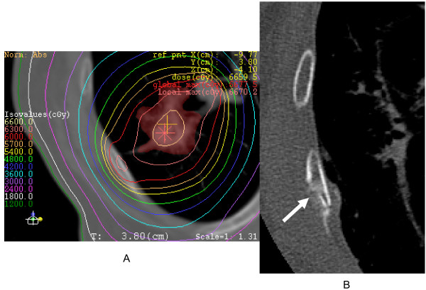
A total of 177 patients who had undergone SRT were prospectively evaluated for periodical follow-up thin-section CT with special attention to chest wall injury. The time at which CT findings of chest wall injury appeared was assessed. Related clinical symptoms were also evaluated.
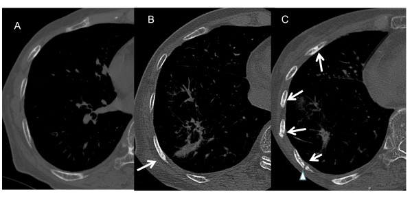
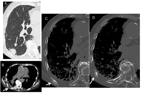
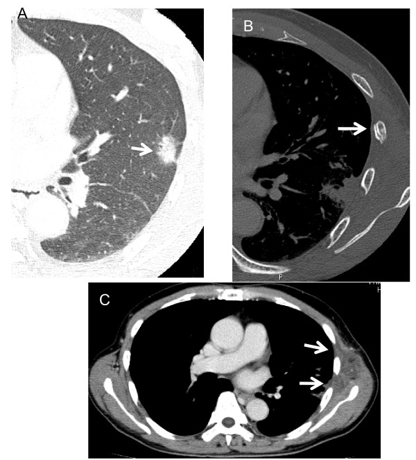
Rib fracture was identified on follow-up CT in 41 patients (23.2%). Rib fractures appeared at a mean of 21.2 months after the completion of SRT (range, 4-58 months). Chest wall edema, thinning of the cortex and osteosclerosis were findings frequently associated with, and tending to precede rib fractures. No patients with rib fracture showed tumors > 16 mm from the adjacent chest wall. Chest wall pain was seen in 18 of 177 patients (10.2%), of whom 14 patients developed rib fracture. No patients complained of Grade 3 or more symptoms.
Rib fracture is frequently seen after SRT for lung cancer on CT, and is often associated with chest wall edema, thinning of the cortex and osteosclerosis. However, related chest wall pain is less frequent and is generally mild if present.
立体定向放疗(SRT)治疗原发性肺癌后,最近有报道称会出现胸壁损伤。然而,其详细的影像学表现尚不清楚。因此,本研究旨在充分描述原发性肺癌 SRT 后 CT 上的胸壁损伤表现、出现时间和频率。
共对 177 例接受 SRT 的患者进行前瞻性评估,定期进行薄层 CT 随访,特别注意胸壁损伤。评估胸壁损伤 CT 表现出现的时间。还评估了相关的临床症状。
41 例(23.2%)患者在随访 CT 上发现肋骨骨折。肋骨骨折出现在 SRT 完成后平均 21.2 个月(范围为 4-58 个月)。胸壁水肿、皮质变薄和骨硬化是常与肋骨骨折相关且倾向于先于肋骨骨折出现的表现。无肋骨骨折患者的肿瘤距相邻胸壁>16mm。177 例患者中有 18 例(10.2%)出现胸痛,其中 14 例发生肋骨骨折。无患者出现 3 级或更高级别的症状。
肺癌 SRT 后 CT 上常可见肋骨骨折,常伴有胸壁水肿、皮质变薄和骨硬化。然而,如果存在,相关的胸痛发生频率较低,且通常较轻。