Department of Chemical Engineering, University of Florida, Gainesville, FL 32611, USA.
Mol Biol Cell. 2011 Dec;22(24):4834-41. doi: 10.1091/mbc.E11-07-0611. Epub 2011 Oct 19.
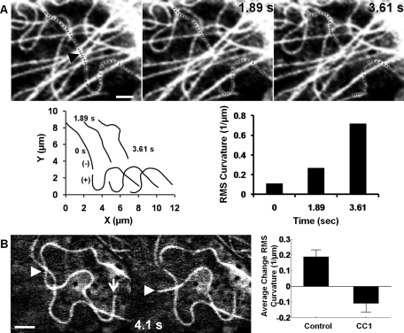
To determine forces on intracellular microtubules, we measured shape changes of individual microtubules following laser severing in bovine capillary endothelial cells. Surprisingly, regions near newly created minus ends increased in curvature following severing, whereas regions near new microtubule plus ends depolymerized without any observable change in shape. With dynein inhibited, regions near severed minus ends straightened rapidly following severing. These observations suggest that dynein exerts a pulling force on the microtubule that buckles the newly created minus end. Moreover, the lack of any observable straightening suggests that dynein prevents lateral motion of microtubules. To explain these results, we developed a model for intracellular microtubule mechanics that predicts the enhanced buckling at the minus end of a severed microtubule. Our results show that microtubule shapes reflect a dynamic force balance in which dynein motor and friction forces dominate elastic forces arising from bending moments. A centrosomal array of microtubules subjected to dynein pulling forces and resisted by dynein friction is predicted to center on the experimentally observed time scale, with or without the pushing forces derived from microtubule buckling at the cell periphery.
为了确定细胞内微管上的力,我们测量了在牛毛细血管内皮细胞中激光切断后单个微管的形状变化。令人惊讶的是,在新创建的微管负端附近的区域在切断后曲率增加,而在新的微管正端附近的区域解聚而没有任何可观察到的形状变化。在抑制动力蛋白后,切断的负端附近的区域在切断后迅速变直。这些观察结果表明,动力蛋白对微管施加拉力,使新创建的微管负端弯曲。此外,没有任何可观察到的变直表明动力蛋白阻止了微管的侧向运动。为了解释这些结果,我们开发了一个细胞内微管力学模型,该模型预测了切断的微管负端的增强弯曲。我们的结果表明,微管的形状反映了一个动态的力平衡,其中动力蛋白马达和摩擦力主导了由弯曲力矩产生的弹性力。一个受动力蛋白拉力作用并受动力蛋白摩擦力抵抗的中心体微管阵列预计将在实验观察到的时间尺度上居中,无论是否存在源自细胞边缘微管弯曲的推压力。