Abdul Q R, Qayum M S, Saradhi M V, Panigrahi M K, Sreedhar V
Department of orthopedics, Sher-I-Kashmir Institute of Medical Sciences MC Hospital, Srinagar, J & K, India.
J Craniovertebr Junction Spine. 2011 Jan;2(1):12-6. doi: 10.4103/0974-8237.85308.
A prospective clinical study of posterior lumbar interbody fusion in grade I and II degenerative spondylolisthesis was conducted between Mar 2007 and Aug 2008.
The objective was to assess the clinicoradiological profile of structural v/s nonstructural graft on intervertebral disc height and its consequences on the low back pain (LBP) assessed by Visual analog score (VAS) score and oswestry disability index (ODI) . This study involved 28 patients.
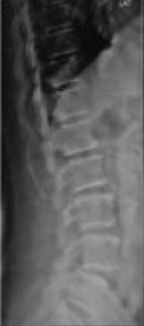
Age of 30-70 years, symptomatic patient with disturbed Activities of daily living (ADL), single-level L4/L5 or L5/S1 grade I or grade II degenerative spondylolisthesis.
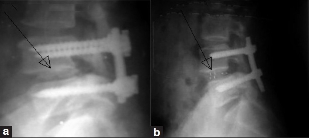
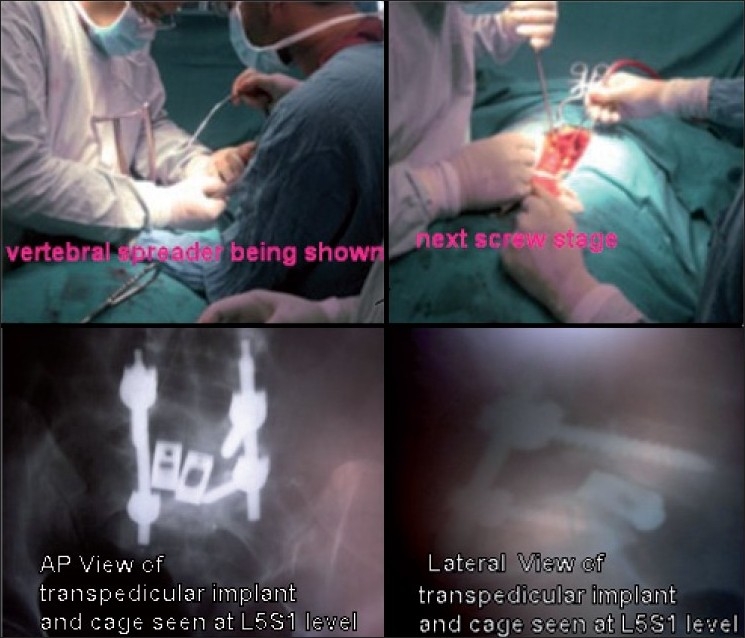
Patients with osteoporosis, recent spondylodiscitis, subchondral sclerosis, visual and cognitive impairment and all other types of spondylolisthesis. All the patients underwent short-segment posterior fixation using CD2 or M8 instrumentation, laminectomy discectomy, reduction and distraction of the involved vertebral space. In 53.5% (n = 15) of the patients, snugly fitted local bone chips were used while in 46.4% (n = 13) of the patients, cage was used. Among the cage group, titanium cage was used in nine (32.1%) and PEEK cages were used in four (14.2%) patients. In one patient, a unilateral PEEK cage was used. The mean follow-up period was 24 months. Among the 28 patients, 67.8% (n = 19) were females and 32.14% (n = 9) were males. 68.24% (n = 18) had L4/L5 and 35.71% (n = 10) had L5/S1 spondylolisthesis. 39.28% (n = 11) were of grade I and 60.71% (n = 17) were of grade II spondylolisthesis.
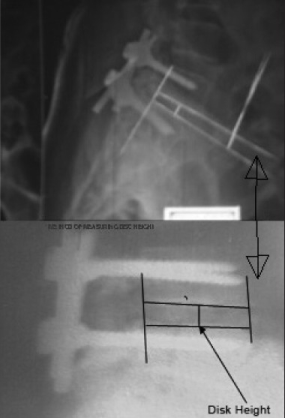
There was a statistically significant correlation (P < 0.012 and P < 0.027) between the change in disc height achieved and the improvement in VAS score in both the graft group and the cage group. The increment in disc height and VAS score was significantly better in the cage group (2 mm ± SD vis-a-vis 7.2 [88%]) than the graft group (1.2 mm ± SD vis-a-vis 5 [62 %]).
2007年3月至2008年8月间对I级和II级退行性椎体滑脱患者进行了腰椎后路椎间融合术的前瞻性临床研究。
评估结构性与非结构性植骨对椎间盘高度的临床放射学特征及其对通过视觉模拟评分(VAS)和奥斯维斯特残疾指数(ODI)评估的下腰痛(LBP)的影响。本研究纳入了28例患者。
年龄30 - 70岁,日常生活活动(ADL)受干扰的有症状患者,单节段L4/L5或L5/S1 I级或II级退行性椎体滑脱。
骨质疏松症患者、近期脊椎椎间盘炎患者、软骨下硬化患者、视力和认知障碍患者以及所有其他类型的椎体滑脱患者。所有患者均采用CD2或M8器械进行短节段后路固定、椎板切除术、椎间盘切除术、受累椎间隙的复位和撑开。53.5%(n = 15)的患者使用紧密贴合的局部骨块,而46.4%(n = 13)的患者使用椎间融合器。在椎间融合器组中,9例(32.1%)患者使用钛合金椎间融合器,4例(14.2%)患者使用聚醚醚酮(PEEK)椎间融合器。1例患者使用单侧PEEK椎间融合器。平均随访期为24个月。28例患者中,67.8%(n = 19)为女性,32.14%(n = 9)为男性。68.24%(n = 18)患者为L4/L5椎体滑脱,35.71%(n = 10)患者为L5/S1椎体滑脱。39.28%(n = 11)为I级椎体滑脱,60.71%(n = 17)为II级椎体滑脱。
植骨组和椎间融合器组中,椎间盘高度变化与VAS评分改善之间存在统计学显著相关性(P < 0.012和P < 0.027)。椎间融合器组的椎间盘高度增加和VAS评分改善明显优于植骨组(分别为2 mm ±标准差对应7.2 [88%]对比1.2 mm ±标准差对应5 [62%])。