Sharma Shweta, Mohanty Sujata, Gupta Deepika, Jassal Manjeet, Agrawal Ashwini K, Tandon Radhika
Department of Ophthalmology, Dr. Rajendra Prasad Centre for Ophthalmic Sciences, All India Institute of Medical Sciences, New Delhi, India.
Mol Vis. 2011;17:2898-910. Epub 2011 Nov 12.
The aim of this study was to develop a synthetic stromal substrate for limbal epithelial cell (LEC) expansion that can serve as a potential alternative substrate to replace human amniotic membrane (HAM).
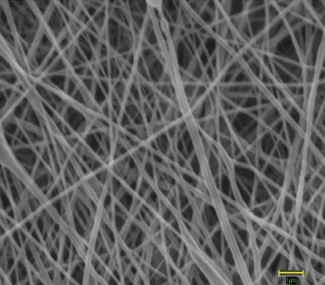
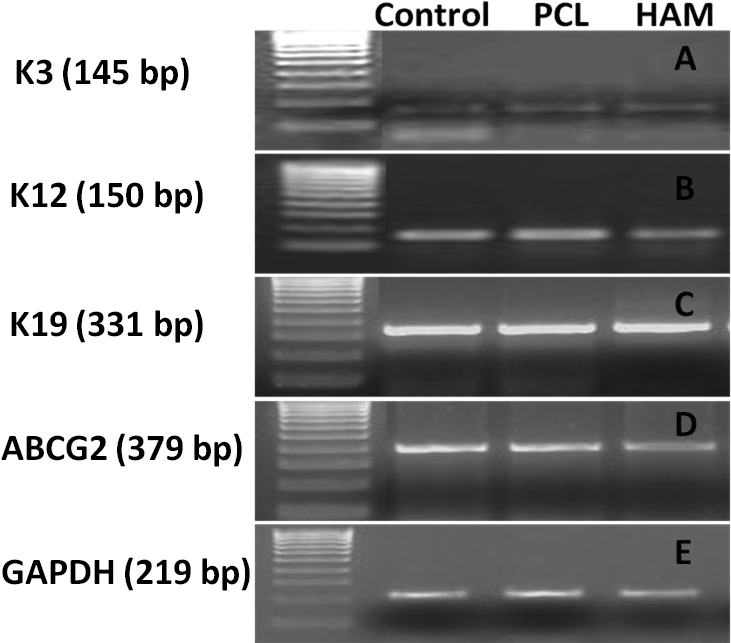
Nanofibers were fabricated using 10% poly-ε-caprolactone (PCL) solution dissolved in trifluoroethanol (TFE) via an electrospinning process. Nanofibers were characterized for surface morphology, wetting ability, pore size, mechanical strength, and optical transparency using scanning electron microscopy (SEM), contact angle measurement, microtensile tester, and UV-Vis spectrophotometer, respectively. The human corneal epithelial (HCE-T) cell line was used to evaluate the biocompatibility of nanofibers based on their phenotypic profile, viability, proliferation, and attachment ability. Subsequently, human LECs were cultivated on biocompatible nanofibers for two weeks and their proliferation capability analyzed using MTT ((3-(4,5-Dimethylthiazol-2-yl)-2,5-diphenyltetrazolium bromide, a yellow tetrazole)) proliferation assay. Immunofluorescent (IF) staining and reverse transcriptase polymerase chain reaction (RT-PCR) were performed to check the molecular marker expression; SEM was used to study the morphology.
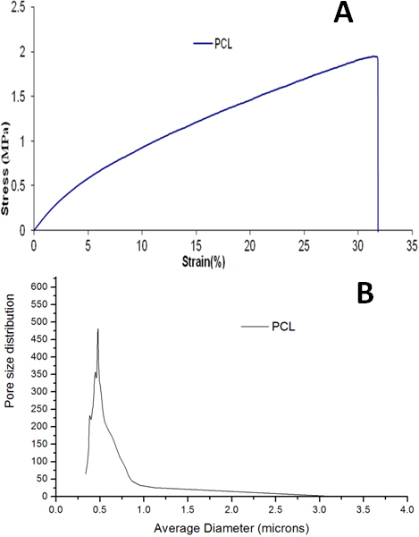
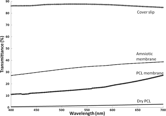
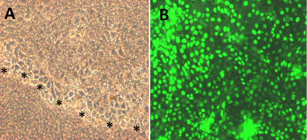
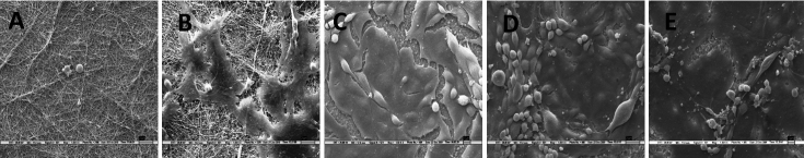
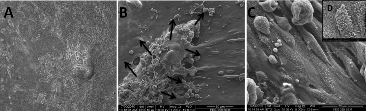
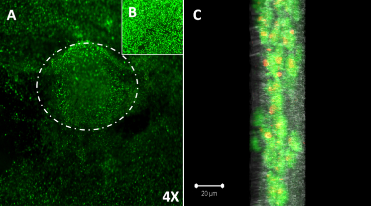
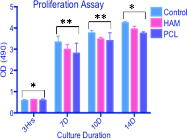
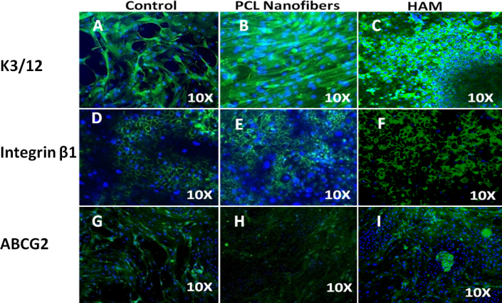
The average fiber diameter of PCL was 132±42 nm. Pore size varied from 0.2 to 4 microns with a porosity of 85%. The tensile strength of the PCL membrane was 1.74±0.18 MPa (Mega Pascal); strain was 30.08±2.66%. The water contact angle was 90°. Biocompatibility results indicated that the polymer surface was highly biocompatible, as HCE-T cells could favorably attach and proliferate on the polymer surface. SEM figures showed that the corneal epithelium was firmly anchored to the polymer surface via a continuous cell sheet and was able to retain a normal corneal phenotype. MTT assay confirmed that cells were metabolically active on nanofibers (p<0.05) and gradually increased in their number for up to two weeks. IF and RT-PCR results revealed no change in the expression profile of LECs grown on nanofibers when compared to those grown on glass coverslips and human amniotic membrane (HAM). Confocal microscopy illustrated that cells infiltrated the nanofibers and successfully formed a three-dimensional (3D) corneal epithelium, which was viable for two weeks.
Electrospun nanofibers provide not only a milieu supporting LEC expansion, but also serve as a useful alternative carrier for ocular surface tissue engineering and could be used as an alternative substrate to HAM.
本研究旨在开发一种用于角膜缘上皮细胞(LEC)扩增的合成基质,作为替代人羊膜(HAM)的潜在基质。
使用溶解在三氟乙醇(TFE)中的10%聚ε-己内酯(PCL)溶液通过静电纺丝工艺制备纳米纤维。分别使用扫描电子显微镜(SEM)、接触角测量仪、微拉伸测试仪和紫外可见分光光度计对纳米纤维的表面形态、润湿性、孔径、机械强度和光学透明度进行表征。使用人角膜上皮(HCE-T)细胞系,根据其表型特征、活力、增殖和附着能力评估纳米纤维的生物相容性。随后,将人LEC在生物相容性纳米纤维上培养两周,并使用MTT((3-(4,5-二甲基噻唑-2-基)-2,5-二苯基四氮唑溴盐,一种黄色四氮唑))增殖试验分析其增殖能力。进行免疫荧光(IF)染色和逆转录聚合酶链反应(RT-PCR)以检查分子标志物表达;使用SEM研究形态。
PCL的平均纤维直径为132±42nm。孔径在0.2至4微米之间变化,孔隙率为85%。PCL膜的拉伸强度为1.74±0.18MPa(兆帕斯卡);应变率为30.08±2.66%。水接触角为90°。生物相容性结果表明,聚合物表面具有高度生物相容性,因为HCE-T细胞能够在聚合物表面良好地附着和增殖。SEM图像显示,角膜上皮通过连续的细胞片牢固地锚定在聚合物表面,并能够保持正常的角膜表型。MTT试验证实细胞在纳米纤维上具有代谢活性(p<0.05),并且其数量在长达两周的时间内逐渐增加。IF和RT-PCR结果显示,与在玻璃盖玻片和人羊膜(HAM)上生长的LEC相比,在纳米纤维上生长的LEC的表达谱没有变化。共聚焦显微镜显示细胞渗入纳米纤维并成功形成三维(3D)角膜上皮,其存活了两周。
静电纺丝纳米纤维不仅提供了支持LEC扩增的环境,而且还可作为眼表组织工程的有用替代载体,并可作为HAM的替代基质。