Department of Periodontology, Faculty of Dentistry, Gazi University, Ankara, Turkey.
Med Oral Patol Oral Cir Bucal. 2012 Jan 1;17(1):e171-7. doi: 10.4317/medoral.17336.
Platelet-rich plasma (PRP) is considered to enhance bone formation especially at early stages of wound healing, depending on the limited and short life-span of platelets and growth factors. The aim of this study was to evaluate efficacy of double-application of PRP (DA-PRP) on bone healing in a rabbit calvarial defect model.
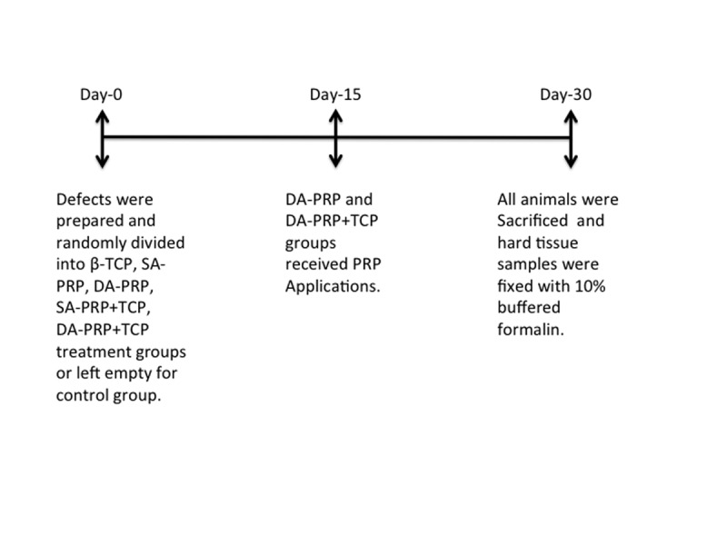
Twenty-eight rabbits, each had two surgically prepared calvarial bone defects (10mm diameter), were included in this study and randomly divided into six groups. Defects (n=56) were treated with single-application of PRP (SA-PRP)(n=10), SA-PRP and beta-tricalciumphosphate (SA-PRP+TCP)(n=10), DA-PRP (n=8), DA-PRP and beta-tricalciumphosphate (DA-PRP+TCP)(n=8), beta-tricalciumphosphate (TCP)(n=10) or left empty (Control)(n=10). Animals were sacrificed at 30 days postoperatively.
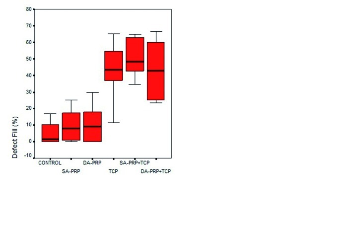
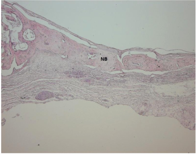
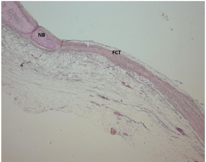
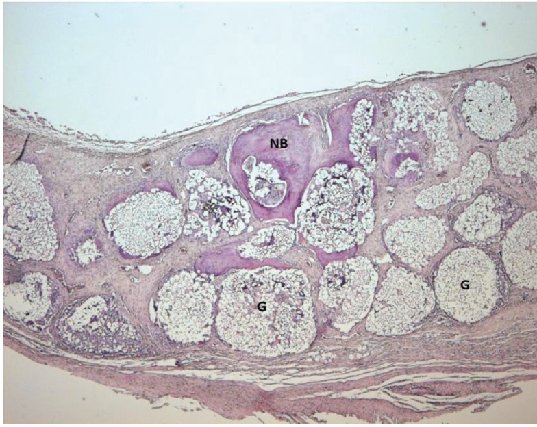
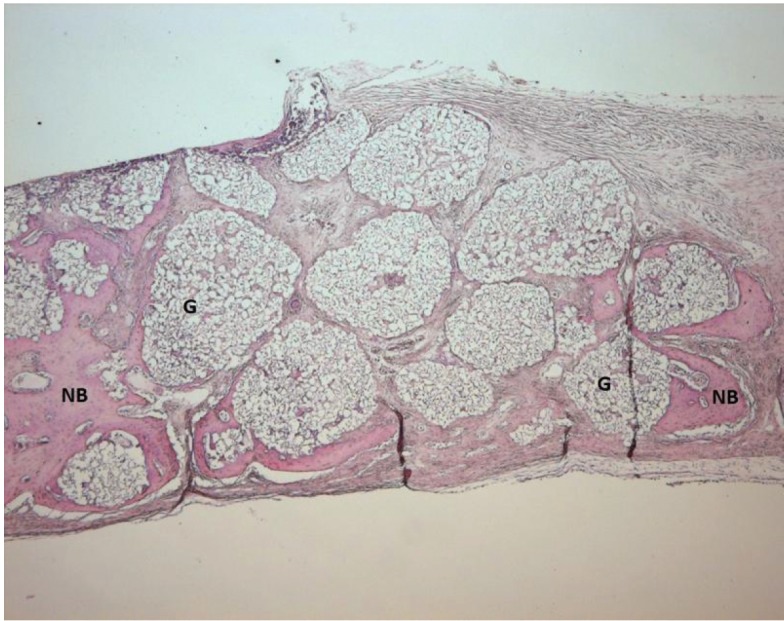
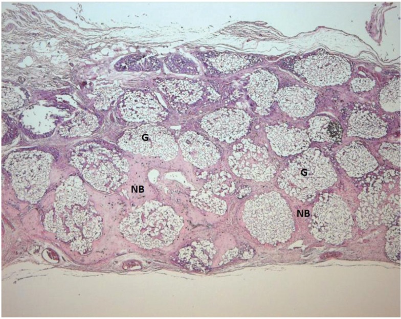
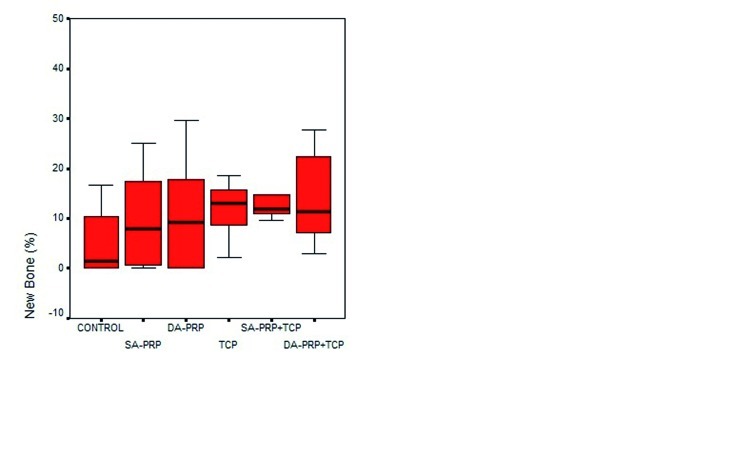
The new bone (NB%) and defect fill (DF%) percentages were calculated from histological slides by image-analyzer software and statistically analysed. All test groups showed higher NB% than control, but differences among all groups were insignificant. The TCP treated groups had significantly higher DF% than groups treated without TCP, however the DF% differences between control, SA-PRP and DA-PRP or TCP, SA-PRP+TCP or DA-PRP+TCP were insignificant.
Although new bone formation was histomorphologically remarkable at double-application PRP groups, statistical analyses of the histomorphometric data revealed no significant difference.
富血小板血浆(PRP)被认为可以增强骨形成,特别是在伤口愈合的早期阶段,这取决于血小板和生长因子的有限和短寿命。本研究的目的是评估双次应用 PRP(DA-PRP)在兔颅骨缺损模型中对骨愈合的疗效。
28 只兔子,每只兔子有两个手术制备的颅骨骨缺损(直径 10mm),被纳入本研究并随机分为六组。用单次应用 PRP(SA-PRP)(n=10)、SA-PRP 和β-磷酸三钙(SA-PRP+TCP)(n=10)、DA-PRP(n=8)、DA-PRP 和β-磷酸三钙(DA-PRP+TCP)(n=8)、β-磷酸三钙(TCP)(n=10)或空白对照(n=10)治疗缺陷(n=56)。动物在手术后 30 天被处死。
通过图像分析软件从组织学切片计算新骨(NB%)和缺损填充(DF%)百分比,并进行统计学分析。所有实验组的 NB%均高于对照组,但组间差异无统计学意义。TCP 治疗组的 DF%明显高于未用 TCP 治疗的组,但对照组、SA-PRP 和 DA-PRP 或 TCP、SA-PRP+TCP 或 DA-PRP+TCP 之间的 DF%差异无统计学意义。
尽管在双次应用 PRP 组中,新骨形成在组织形态学上是显著的,但组织形态计量学数据的统计分析显示无显著差异。