Department of Molecular Structural Biology, Max Planck Institute of Biochemistry, Martinsried, Germany.
PLoS Pathog. 2011 Dec;7(12):e1002406. doi: 10.1371/journal.ppat.1002406. Epub 2011 Dec 15.
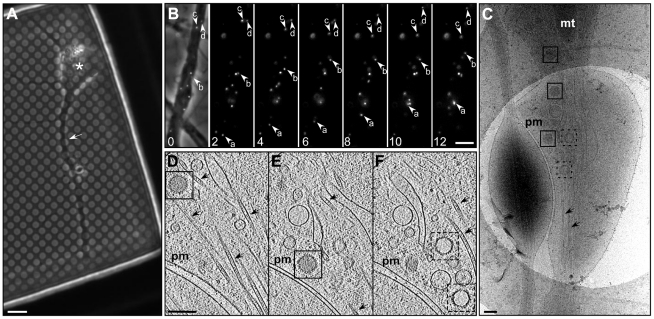
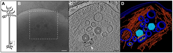
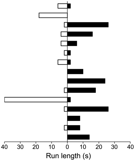
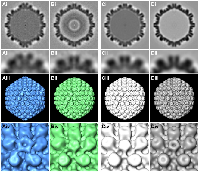
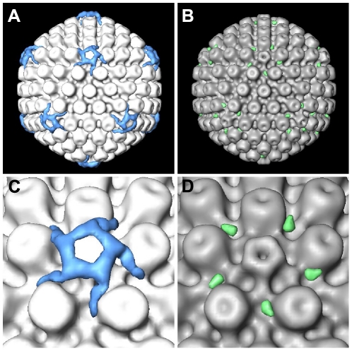
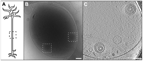
During herpes simplex virus 1 (HSV1) egress in neurons, viral particles travel from the neuronal cell body along the axon towards the synapse. Whether HSV1 particles are transported as enveloped virions as proposed by the 'married' model or as non-enveloped capsids suggested by the 'separate' model is controversial. Specific viral proteins may form a recruitment platform for microtubule motors that catalyze such transport. However, their subviral location has remained elusive. Here we established a system to analyze herpesvirus egress by cryo electron tomography. At 16 h post infection, we observed intra-axonal transport of progeny HSV1 viral particles in dissociated hippocampal neurons by live-cell fluorescence microscopy. Cryo electron tomography of frozen-hydrated neurons revealed that most egressing capsids were transported independently of the viral envelope. Unexpectedly, we found not only DNA-containing capsids (cytosolic C-capsids), but also capsids lacking DNA (cytosolic A-/B-capsids) in mid-axon regions. Subvolume averaging revealed lower amounts of tegument on cytosolic A-/B-capsids than on C-capsids. Nevertheless, all capsid types underwent active axonal transport. Therefore, even few tegument proteins on the capsid vertices seemed to suffice for transport. Secondary envelopment of capsids was observed at axon terminals. On their luminal face, the enveloping vesicles were studded with typical glycoprotein-like spikes. Furthermore, we noted an accretion of tegument density at the concave cytosolic face of the vesicle membrane in close proximity to the capsids. Three-dimensional analysis revealed that these assembly sites lacked cytoskeletal elements, but that filamentous actin surrounded them and formed an assembly compartment. Our data support the 'separate model' for HSV1 egress, i.e. progeny herpes viruses being transported along axons as subassemblies and not as complete virions within transport vesicles.
在单纯疱疹病毒 1 (HSV1) 从神经元中出芽时,病毒颗粒沿着轴突从神经元细胞体向突触移动。HSV1 颗粒是作为“已婚”模型所提出的包膜病毒,还是作为“分离”模型所提出的无包膜衣壳进行运输,这仍然存在争议。特定的病毒蛋白可能形成微管马达的募集平台,从而促进这种运输。然而,它们的亚病毒位置仍然难以捉摸。在这里,我们建立了一个通过低温电子断层扫描分析疱疹病毒出芽的系统。在感染后 16 小时,我们通过活细胞荧光显微镜观察到分离的海马神经元中新生 HSV1 病毒颗粒的轴内运输。冷冻水合神经元的低温电子断层扫描显示,大多数出芽的衣壳是独立于病毒包膜进行运输的。出乎意料的是,我们不仅在中间轴突区域发现了含有 DNA 的衣壳(细胞质 C-衣壳),还发现了不含 DNA 的衣壳(细胞质 A-/B-衣壳)。子体积平均化显示,细胞质 A-/B-衣壳上的被膜蛋白比 C-衣壳上的要少。尽管如此,所有衣壳类型都经历了活跃的轴突运输。因此,即使衣壳顶点上的少量被膜蛋白似乎足以进行运输。衣壳的二次包被发生在轴突末端。在囊泡的腔面,被膜上点缀着典型的糖蛋白样刺突。此外,我们还注意到囊泡膜的胞质面有被膜蛋白密度的堆积,与衣壳紧密相邻。三维分析显示,这些组装部位缺乏细胞骨架成分,但丝状肌动蛋白围绕着它们并形成一个组装隔室。我们的数据支持 HSV1 出芽的“分离模型”,即子代疱疹病毒作为亚组装体沿着轴突运输,而不是作为完整的病毒颗粒在运输囊泡内运输。