• 文献检索
  • 文档翻译
  • 深度研究
  • 学术资讯
  • Suppr Zotero 插件Zotero 插件
  • 邀请有礼
  • 套餐&价格
  • 历史记录
应用&插件
Suppr Zotero 插件Zotero 插件浏览器插件Mac 客户端Windows 客户端微信小程序
定价
高级版会员购买积分包购买API积分包
服务
文献检索文档翻译深度研究API 文档MCP 服务
关于我们
关于 Suppr公司介绍联系我们用户协议隐私条款
关注我们

Suppr 超能文献

核心技术专利:CN118964589B侵权必究
粤ICP备2023148730 号-1Suppr @ 2026

文献检索

告别复杂PubMed语法,用中文像聊天一样搜索,搜遍4000万医学文献。AI智能推荐,让科研检索更轻松。

立即免费搜索

文件翻译

保留排版,准确专业,支持PDF/Word/PPT等文件格式,支持 12+语言互译。

免费翻译文档

深度研究

AI帮你快速写综述,25分钟生成高质量综述,智能提取关键信息,辅助科研写作。

立即免费体验

机械微环境通过冷冻电子断层扫描来调节轴突直径。

The Mechanical Microenvironment Regulates Axon Diameters Visualized by Cryo-Electron Tomography.

机构信息

Ohio State Biochemistry Graduate Program, The Ohio State University, Columbus, OH 43210, USA.

Department of Biological Chemistry and Pharmacology, The Ohio State University, Columbus, OH 43210, USA.

出版信息

Cells. 2022 Aug 15;11(16):2533. doi: 10.3390/cells11162533.

DOI:10.3390/cells11162533
PMID:36010609
原文链接:https://pmc.ncbi.nlm.nih.gov/articles/PMC9406316/
Abstract

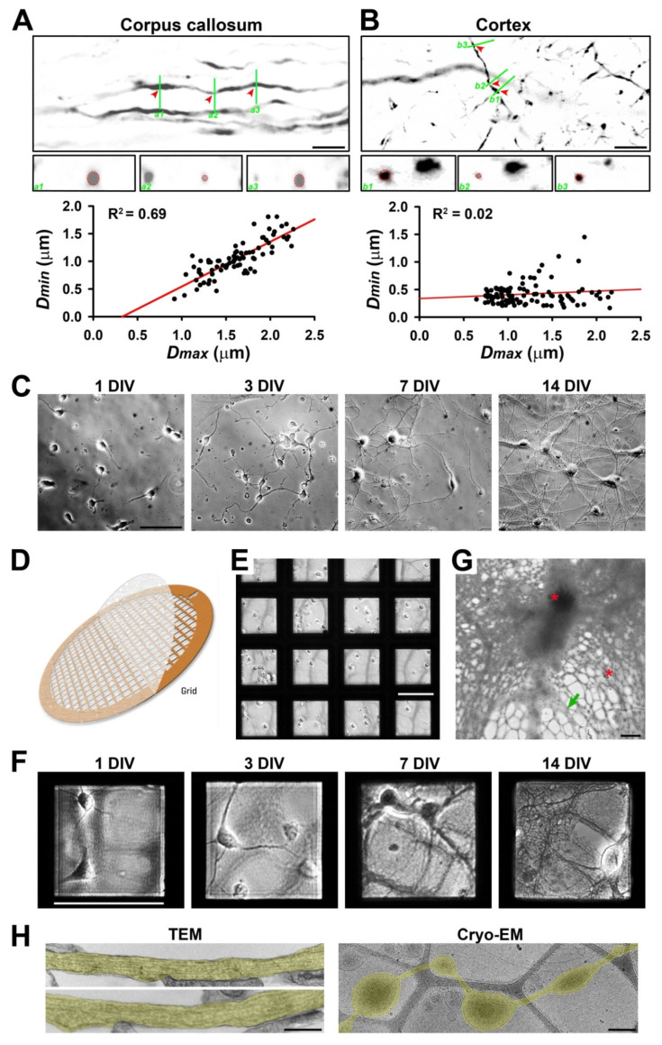
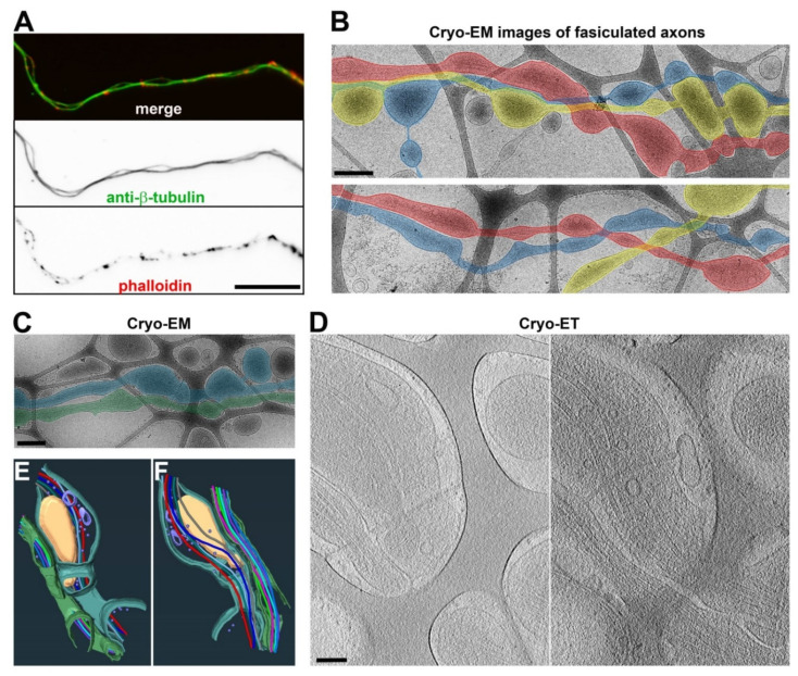
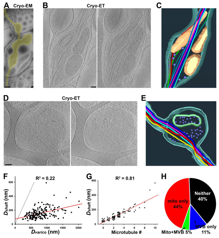
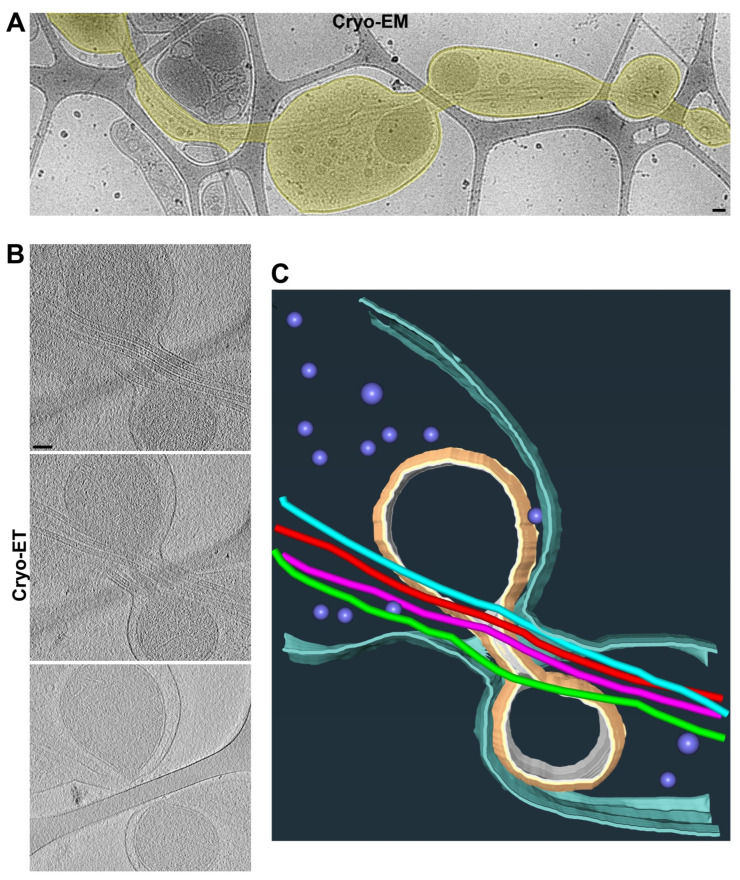
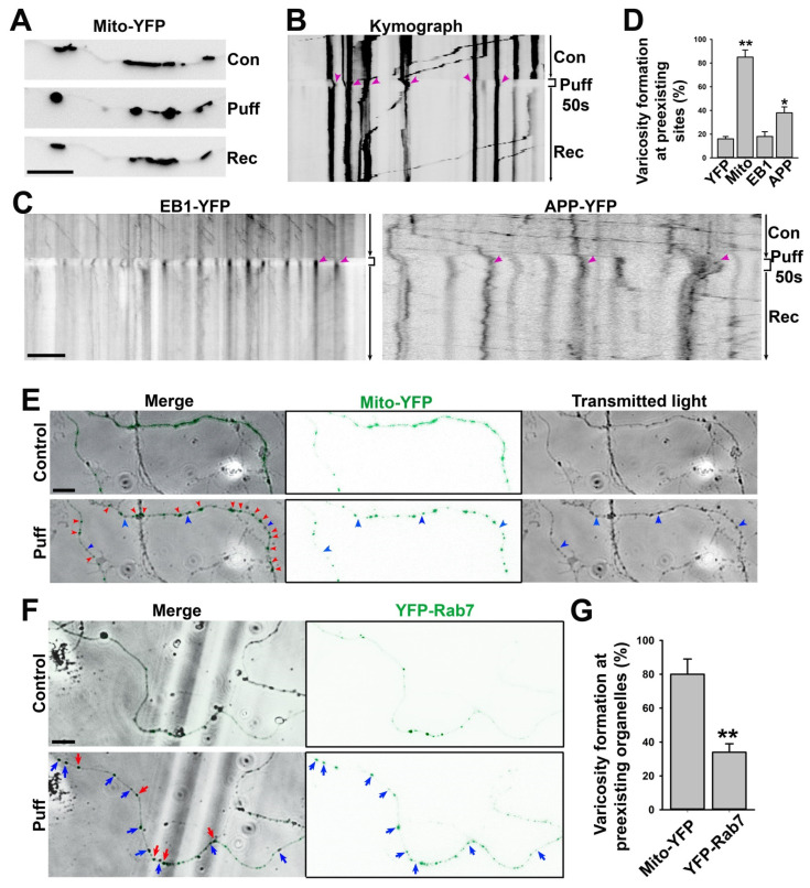
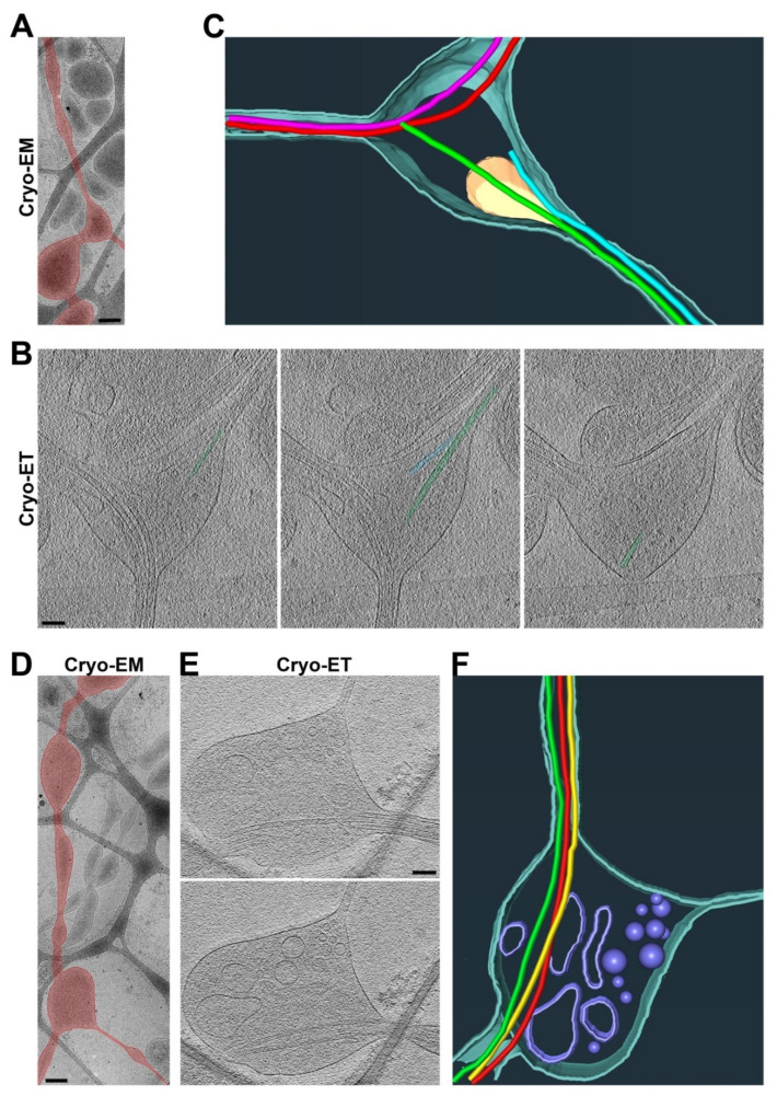
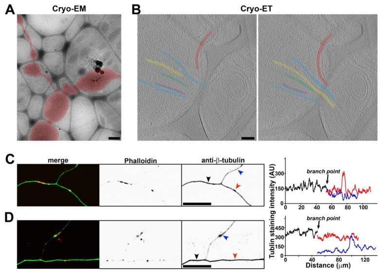
Axonal varicosities or swellings are enlarged structures along axon shafts and profoundly affect action potential propagation and synaptic transmission. These structures, which are defined by morphology, are highly heterogeneous and often investigated concerning their roles in neuropathology, but why they are present in the normal brain remains unknown. Combining confocal microscopy and cryo-electron tomography (Cryo-ET) with in vivo and in vitro systems, we report that non-uniform mechanical interactions with the microenvironment can lead to 10-fold diameter differences within an axon of the central nervous system (CNS). In the brains of adult Thy1-YFP transgenic mice, individual axons in the cortex displayed significantly higher diameter variation than those in the corpus callosum. When being cultured on lacey carbon film-coated electron microscopy (EM) grids, CNS axons formed varicosities exclusively in holes and without microtubule (MT) breakage, and they contained mitochondria, multivesicular bodies (MVBs), and/or vesicles, similar to the axonal varicosities induced by mild fluid puffing. Moreover, enlarged axon branch points often contain MT free ends leading to the minor branch. When the axons were fasciculated by mimicking in vivo axonal bundles, their varicosity levels reduced. Taken together, our results have revealed the extrinsic regulation of the three-dimensional ultrastructures of central axons by the mechanical microenvironment under physiological conditions.

摘要

轴突膨体或肿胀是沿着轴突轴的扩大结构,对动作电位的传播和突触传递有深远影响。这些结构根据形态定义,高度异质,并且经常针对其在神经病理学中的作用进行研究,但它们在正常大脑中存在的原因仍然未知。通过将共聚焦显微镜和冷冻电子断层扫描(Cryo-ET)与体内和体外系统相结合,我们报告称,与微环境的非均匀机械相互作用可导致中枢神经系统(CNS)轴突内的直径差异达到 10 倍。在成年 Thy1-YFP 转基因小鼠的大脑中,皮质中的单个轴突的直径变化明显高于胼胝体中的轴突。当在带有 lacey 碳膜的电子显微镜(EM)网格上培养时,CNS 轴突仅在孔中形成膨体,而不会发生微管(MT)断裂,并且它们包含线粒体、多泡体(MVB)和/或囊泡,与通过温和流体脉冲诱导的轴突膨体相似。此外,扩大的轴突分支点通常包含 MT 无末端,导致小分支。当通过模拟体内轴突束来聚集轴突时,它们的膨体水平降低。总之,我们的结果揭示了在生理条件下机械微环境对中枢轴突的三维超微结构的外在调节。

https://cdn.ncbi.nlm.nih.gov/pmc/blobs/f11a/9406316/08f4a3a309e1/cells-11-02533-g007.jpg
https://cdn.ncbi.nlm.nih.gov/pmc/blobs/f11a/9406316/4d6220b8c565/cells-11-02533-g001.jpg
https://cdn.ncbi.nlm.nih.gov/pmc/blobs/f11a/9406316/fb62ef54efe9/cells-11-02533-g002.jpg
https://cdn.ncbi.nlm.nih.gov/pmc/blobs/f11a/9406316/53437f428093/cells-11-02533-g003.jpg
https://cdn.ncbi.nlm.nih.gov/pmc/blobs/f11a/9406316/6d31cdca7ebf/cells-11-02533-g004.jpg
https://cdn.ncbi.nlm.nih.gov/pmc/blobs/f11a/9406316/695c9d5f5c65/cells-11-02533-g005.jpg
https://cdn.ncbi.nlm.nih.gov/pmc/blobs/f11a/9406316/3212f46822ce/cells-11-02533-g006.jpg
https://cdn.ncbi.nlm.nih.gov/pmc/blobs/f11a/9406316/08f4a3a309e1/cells-11-02533-g007.jpg

相似文献

1
The Mechanical Microenvironment Regulates Axon Diameters Visualized by Cryo-Electron Tomography.机械微环境通过冷冻电子断层扫描来调节轴突直径。
Cells. 2022 Aug 15;11(16):2533. doi: 10.3390/cells11162533.
2
Partial interruption of axonal transport due to microtubule breakage accounts for the formation of periodic varicosities after traumatic axonal injury.轴突运输的部分中断是由于微管断裂,这解释了创伤性轴索损伤后周期性泡状突起的形成。
Exp Neurol. 2012 Jan;233(1):364-72. doi: 10.1016/j.expneurol.2011.10.030. Epub 2011 Nov 4.
3
Immediate induction of varicosities by transverse compression but not uniaxial stretch in axon mechanosensation.轴突机械感觉中,横向压缩而非单轴拉伸可即刻诱导曲张。
Acta Neuropathol Commun. 2022 Jan 24;10(1):7. doi: 10.1186/s40478-022-01309-8.
4
Unrestrained growth of correctly oriented microtubules instructs axonal microtubule orientation.正确取向的微管的无约束生长指导着轴突微管的取向。
Elife. 2022 Oct 10;11:e77608. doi: 10.7554/eLife.77608.
5
Morphology of mitochondria in spatially restricted axons revealed by cryo-electron tomography.冷冻电镜断层成像术揭示了空间受限轴突中线粒体的形态。
PLoS Biol. 2018 Sep 17;16(9):e2006169. doi: 10.1371/journal.pbio.2006169. eCollection 2018 Sep.
6
Mechanisms of axonal spheroid formation in central nervous system Wallerian degeneration.中枢神经系统 Wallerian 变性中轴突球体形成的机制。
J Neuropathol Exp Neurol. 2010 May;69(5):455-72. doi: 10.1097/NEN.0b013e3181da84db.
7
Cryo electron tomography of herpes simplex virus during axonal transport and secondary envelopment in primary neurons.单纯疱疹病毒在原发性神经元中的轴突运输和二次包膜形成过程中的冷冻电子断层扫描。
PLoS Pathog. 2011 Dec;7(12):e1002406. doi: 10.1371/journal.ppat.1002406. Epub 2011 Dec 15.
8
Rapid and Reversible Development of Axonal Varicosities: A New Form of Neural Plasticity.轴突膨体的快速可逆发展:一种新的神经可塑性形式。
Front Mol Neurosci. 2021 Feb 3;14:610857. doi: 10.3389/fnmol.2021.610857. eCollection 2021.
9
Mechanical breaking of microtubules in axons during dynamic stretch injury underlies delayed elasticity, microtubule disassembly, and axon degeneration.在动态拉伸损伤过程中,轴突中的微管机械断裂是延迟弹性、微管解体和轴突退化的基础。
FASEB J. 2010 May;24(5):1401-10. doi: 10.1096/fj.09-142844. Epub 2009 Dec 17.
10
CLARITY reveals a more protracted temporal course of axon swelling and disconnection than previously described following traumatic brain injury.CLARITY 揭示了创伤性脑损伤后轴突肿胀和分离的时间过程比以前描述的更为持久。
Brain Pathol. 2019 May;29(3):437-450. doi: 10.1111/bpa.12677. Epub 2018 Dec 27.

引用本文的文献

1
Precision Recovery After Spinal Cord Injury: Integrating CRISPR Technologies, AI-Driven Therapeutics, Single-Cell Omics, and System Neuroregeneration.脊髓损伤后的精准恢复:整合CRISPR技术、人工智能驱动的疗法、单细胞组学和系统神经再生
Int J Mol Sci. 2025 Jul 20;26(14):6966. doi: 10.3390/ijms26146966.
2
The Roles of Exosomes in Anti-Cancer Drugs.外泌体在抗癌药物中的作用。
Cancer Med. 2025 May;14(9):e70897. doi: 10.1002/cam4.70897.
3
Membrane mechanics dictate axonal pearls-on-a-string morphology and function.膜力学决定轴突上珍珠串样形态和功能。

本文引用的文献

1
Immediate induction of varicosities by transverse compression but not uniaxial stretch in axon mechanosensation.轴突机械感觉中,横向压缩而非单轴拉伸可即刻诱导曲张。
Acta Neuropathol Commun. 2022 Jan 24;10(1):7. doi: 10.1186/s40478-022-01309-8.
2
A cryo-ET survey of microtubules and intracellular compartments in mammalian axons.哺乳动物轴突中微管和细胞内隔室的冷冻电镜调查。
J Cell Biol. 2022 Feb 7;221(2). doi: 10.1083/jcb.202103154. Epub 2021 Dec 8.
3
Electron cryo-tomography reveals the subcellular architecture of growing axons in human brain organoids.
Nat Neurosci. 2025 Jan;28(1):49-61. doi: 10.1038/s41593-024-01813-1. Epub 2024 Dec 2.
4
Actomyosin-II protects axons from degeneration induced by mild mechanical stress.肌球蛋白 II 保护轴突免受轻度机械应激诱导的变性。
J Cell Biol. 2024 Aug 5;223(8). doi: 10.1083/jcb.202206046. Epub 2024 May 7.
5
Handling Difficult Cryo-ET Samples: A Study with Primary Neurons from Drosophila melanogaster.处理困难的冷冻电镜样品:使用黑腹果蝇原代神经元进行的研究。
Microsc Microanal. 2023 Dec 21;29(6):2127-2148. doi: 10.1093/micmic/ozad125.
6
An experimental platform for stochastic analyses of single serotonergic fibers in the mouse brain.用于小鼠大脑中单个血清素能纤维随机分析的实验平台。
Front Neurosci. 2023 Oct 6;17:1241919. doi: 10.3389/fnins.2023.1241919. eCollection 2023.
7
High-resolution spatiotemporal analysis of single serotonergic axons in an system.在一个系统中对单个血清素能轴突进行高分辨率时空分析。
Front Neurosci. 2022 Oct 24;16:994735. doi: 10.3389/fnins.2022.994735. eCollection 2022.
电子冷冻断层成像揭示了人类脑类器官中生长轴突的亚细胞结构。
Elife. 2021 Oct 26;10:e70269. doi: 10.7554/eLife.70269.
4
Cytoskeleton and Membrane Organization at Axon Branches.轴突分支处的细胞骨架与膜组织
Front Cell Dev Biol. 2021 Sep 1;9:707486. doi: 10.3389/fcell.2021.707486. eCollection 2021.
5
In situ architecture of neuronal α-Synuclein inclusions.神经元α-突触核蛋白包涵体的原位结构。
Nat Commun. 2021 Apr 14;12(1):2110. doi: 10.1038/s41467-021-22108-0.
6
Rapid and Reversible Development of Axonal Varicosities: A New Form of Neural Plasticity.轴突膨体的快速可逆发展:一种新的神经可塑性形式。
Front Mol Neurosci. 2021 Feb 3;14:610857. doi: 10.3389/fnmol.2021.610857. eCollection 2021.
7
Putting the axonal periodic scaffold in order.整理轴突周期性支架。
Curr Opin Neurobiol. 2021 Aug;69:33-40. doi: 10.1016/j.conb.2020.12.015. Epub 2021 Jan 12.
8
Mesophasic organization of GABA receptors in hippocampal inhibitory synapses.γ-氨基丁酸受体在海马抑制性突触中的中间相组织。
Nat Neurosci. 2020 Dec;23(12):1589-1596. doi: 10.1038/s41593-020-00729-w. Epub 2020 Nov 2.
9
In situ cryo-electron tomography reveals filamentous actin within the microtubule lumen.原位冷冻电子断层成像揭示了微管腔中的丝状肌动蛋白。
J Cell Biol. 2020 Sep 7;219(9). doi: 10.1083/jcb.201911154.
10
Effects of Focal Axonal Swelling Level on the Action Potential Signal Transmission.轴突局限性肿胀程度对动作电位信号传递的影响。
J Comput Neurosci. 2020 Aug;48(3):253-263. doi: 10.1007/s10827-020-00750-9.