Respiratory Department, Xiangya Hospital, Central South University, Changsha, Hunan, China.
PLoS One. 2011;6(12):e27113. doi: 10.1371/journal.pone.0027113. Epub 2011 Dec 28.
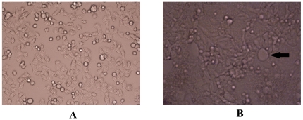
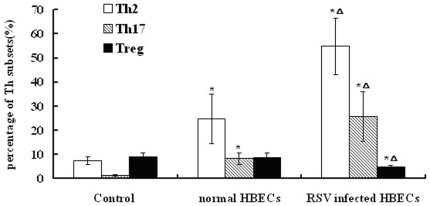
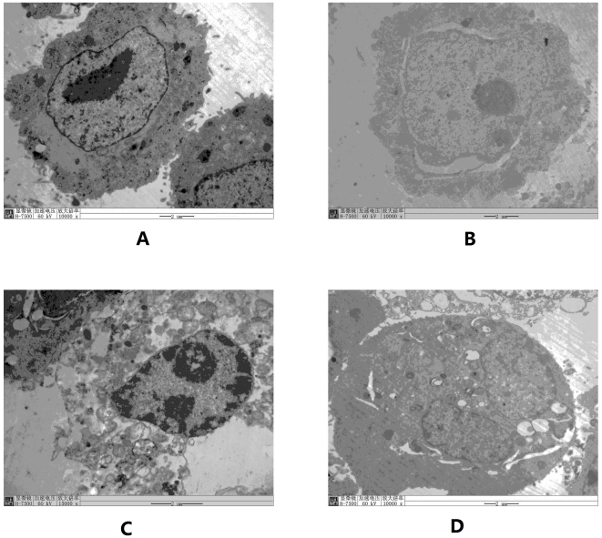
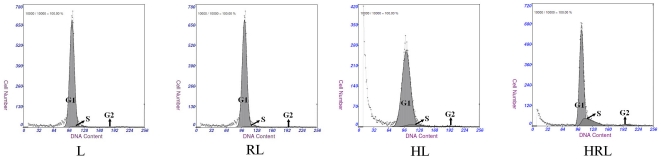
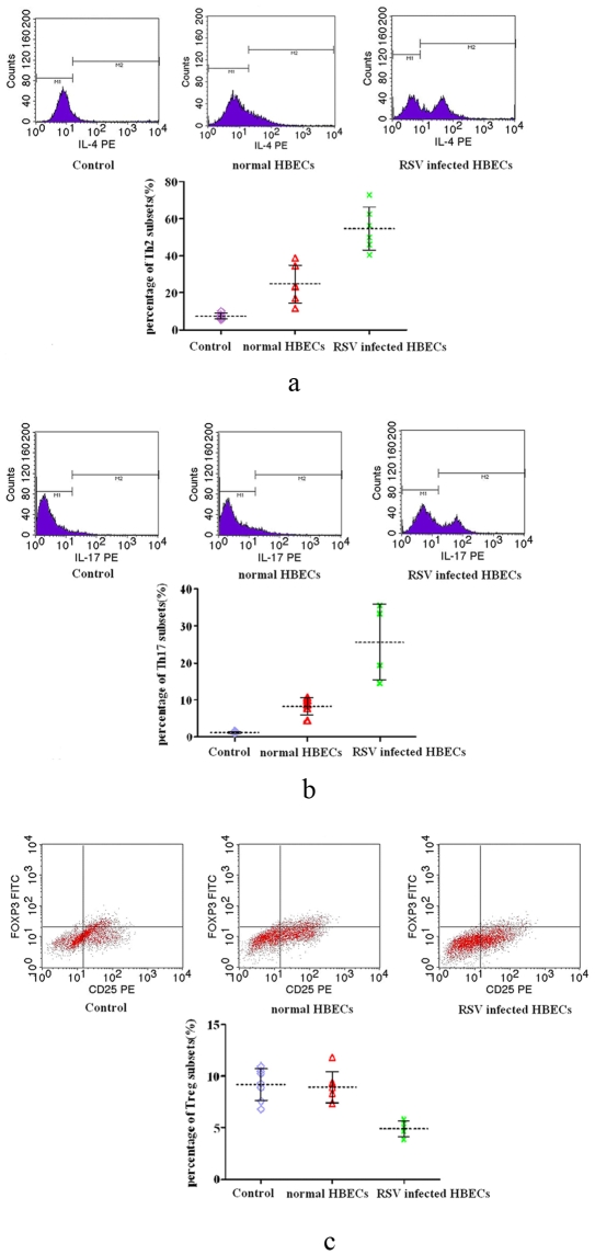
Respiratory syncytial virus (RSV) preferentially infects airway epithelial cells,which might be responsible for susceptibility to asthma; however, the underlying mechanism is not clear. This study determined the activation of lymphocytes and drift of helper T (Th) subsets induced by RSV-infected human bronchial epithelial cells (HBECs) in vitro. HBECs had prolonged infection with RSV, and lymphocytes isolated from human peripheral blood were co-cultured with RSV-infected HBECs. Four groups were established, as follows: lymphocytes (group L); lymphocytes infected with RSV (group RL); co-culture of lymphocytes with non-infected HBECs (group HL); and co-culture of lymphocytes with infected HBECs (group HRL). After co-culture with HBECs for 24 hours, lymphocytes were collected and the following were determined in the 4 groups: cell cycle status; apoptosis rate; and concentrations of IL-4, IFN-γ, and IL-17 in the supernatants. Cell cycle analysis for lymphocytes showed a significant increase in S phase cells, a decrease in G1 phase cells, and a higher apoptosis rate in group HRL compared with the other three groups. In group HRL, the levels of IL-4, IFN-γ, and IL-17 in supernatants were also higher than the other three groups. For further study, lymphocytes were individually treated with supernatants from non-infected and RSV-infected HBECs for 24 h. We showed that supernatants from RSV-infected HBECs induced the differentiation of Th2 and Th17 subsets, and suppressed the differentiation of Treg subsets. Our results showed that HBECs with prolonged RSV infection can induce lymphocyte proliferation and apoptosis, and enhance the release of cytokines by lymphocytes. Moreover, subset drift might be caused by RSV-infected HBECs.
呼吸道合胞病毒(RSV)优先感染气道上皮细胞,这可能是导致哮喘易感性的原因;然而,其潜在机制尚不清楚。本研究旨在确定 RSV 感染的人支气管上皮细胞(HBEC)体外诱导淋巴细胞激活和辅助性 T(Th)亚群漂移的情况。HBEC 被 RSV 持续感染,人类外周血分离的淋巴细胞与 RSV 感染的 HBEC 共培养。建立了 4 个组,如下所示:淋巴细胞(组 L);感染 RSV 的淋巴细胞(组 RL);未感染 HBEC 的淋巴细胞共培养(组 HL);感染 HBEC 的淋巴细胞共培养(组 HRL)。与 HBEC 共培养 24 小时后,收集淋巴细胞,并在 4 个组中测定以下指标:细胞周期状态;细胞凋亡率;上清液中 IL-4、IFN-γ 和 IL-17 的浓度。淋巴细胞的细胞周期分析显示,与其他 3 个组相比,组 HRL 的 S 期细胞显著增加,G1 期细胞减少,细胞凋亡率更高。在组 HRL 中,上清液中 IL-4、IFN-γ 和 IL-17 的水平也高于其他 3 个组。为了进一步研究,将淋巴细胞分别用未感染和 RSV 感染的 HBEC 的上清液处理 24 小时。结果表明,RSV 感染的 HBEC 上清液诱导 Th2 和 Th17 亚群的分化,并抑制 Treg 亚群的分化。我们的结果表明,长期 RSV 感染的 HBEC 可诱导淋巴细胞增殖和凋亡,并增强淋巴细胞释放细胞因子。此外,亚群漂移可能是由 RSV 感染的 HBEC 引起的。