Shoklo Malaria Research Unit, PO Box 46, Mae Sot, Tak 63110, Thailand.
Malar J. 2012 Jan 5;11:5. doi: 10.1186/1475-2875-11-5.
The presence of malaria parasites and histopathological changes in the placenta are associated with a reduction in birth weight, principally due to intrauterine growth restriction. The aim of this study was to examine the feasibility of studying early pregnancy placental volumes using three-dimensional (3D) ultrasound in a malaria endemic area, as a small volume in the second trimester may be an indicator of intra-uterine growth restriction and placental insufficiency.
Placenta volumes were acquired using a portable ultrasound machine and a 3D ultrasound transducer and estimated using the Virtual Organ Computer-aided AnaLysis (VOCAL) image analysis software package. Intra-observer reliability and limits of agreement of the placenta volume measurements were calculated. Polynomial regression models for the mean and standard deviation as a function of gestational age for the placental volumes of uninfected women were created and tested. Based on these equations each measurement was converted into a z -score. The z-scores of the placental volumes of malaria infected and uninfected women were then compared.
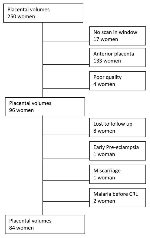
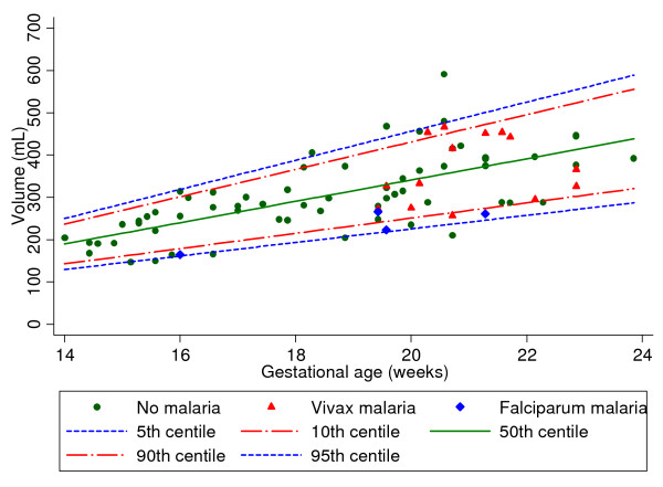
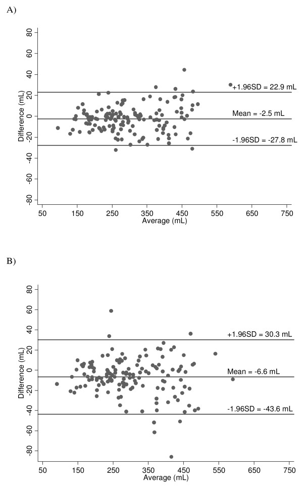
Eighty-four women (uninfected = 65; infected = 19) with a posterior placenta delivered congenitally normal, live born, single babies. The mean placental volumes in the uninfected women were modeled to fit 5th, 10th, 50th, 90th and 95th centiles for 14-24 weeks' gestation. Most placenta volumes in the infected women were below the 50th centile for gestational age; most of those with Plasmodium falciparum were below the 10th centile. The 95% intra-observer limits of agreement for first and second measurements were ± 37.0 mL and ± 25.4 mL at 30 degrees and 15 degrees rotation respectively.
The new technique of 3D ultrasound volumetry of the placenta may be useful to improve our understanding of the pathophysiological constraints on foetal growth caused by malaria infection in early pregnancy.
胎盘内疟原虫的存在和组织病理学变化与出生体重减轻有关,主要是由于宫内生长受限。本研究旨在检查在疟疾流行地区使用三维(3D)超声检查早期妊娠胎盘体积的可行性,因为中期体积较小可能是宫内生长受限和胎盘功能不全的指标。
使用便携式超声机和 3D 超声换能器获取胎盘体积,并使用虚拟器官计算机辅助分析(VOCAL)图像分析软件包进行估计。计算了胎盘体积测量的观察者内可靠性和一致性界限。为未感染妇女的胎盘体积建立了妊娠龄的均数和标准差的多项式回归模型,并进行了测试。基于这些方程,每个测量值都转换为 z 分数。然后比较感染和未感染疟疾妇女的胎盘体积 z 分数。
84 名妇女(未感染= 65;感染= 19)的后胎盘分娩出正常的先天性、活产、单胎婴儿。未感染妇女的平均胎盘体积被建模以适合 14-24 周妊娠的第 5、第 10、第 50、第 90 和第 95 百分位数。大多数感染妇女的胎盘体积低于妊娠龄的第 50 百分位数;大多数感染恶性疟原虫的胎盘体积低于第 10 百分位数。第一次和第二次测量的 95%观察者内一致性界限分别为 30 度和 15 度旋转时的±37.0mL 和±25.4mL。
胎盘 3D 超声体积测量的新技术可能有助于更好地理解早孕感染疟疾对胎儿生长的病理生理限制。