Department of Physics, University of Wisconsin-Milwaukee, Milwaukee, WI 53211, USA.
J Synchrotron Radiat. 2012 Mar;19(Pt 2):264-73. doi: 10.1107/S090904951105549X. Epub 2012 Jan 18.
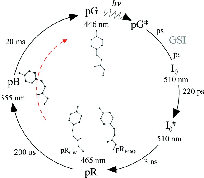
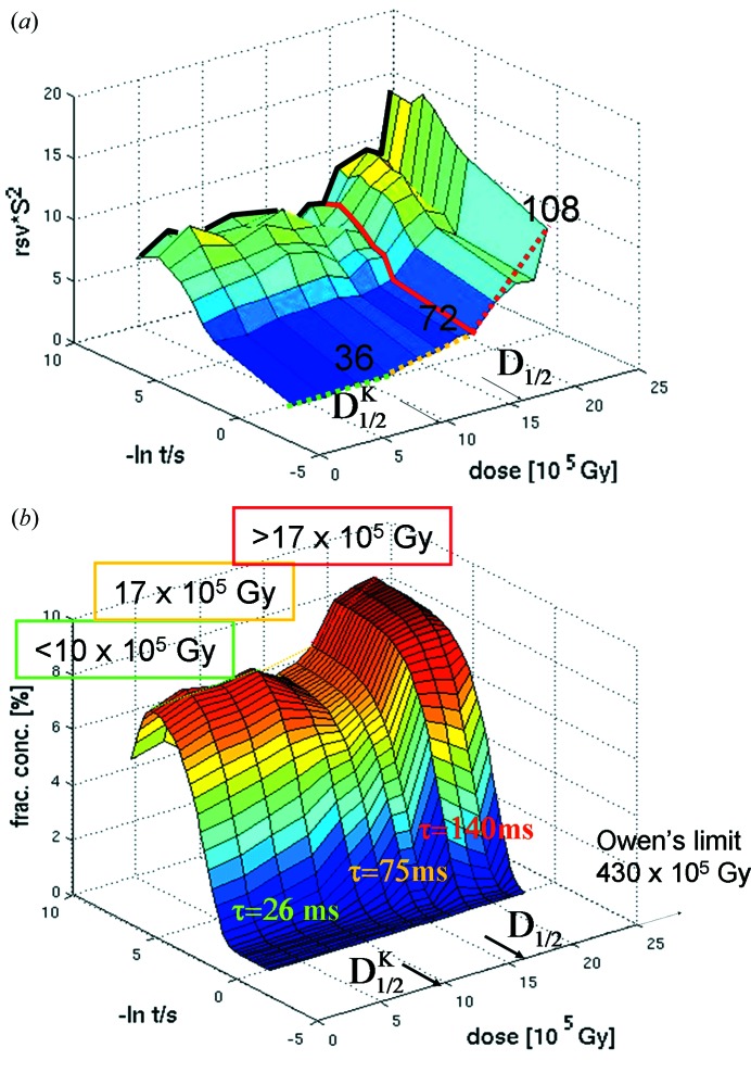
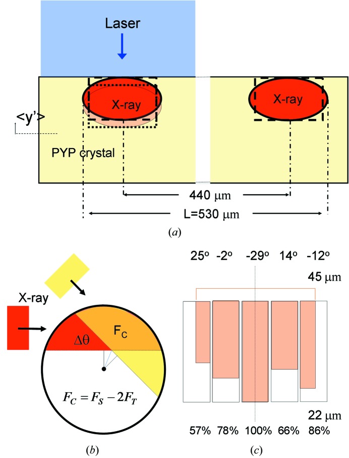
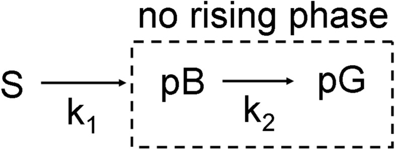
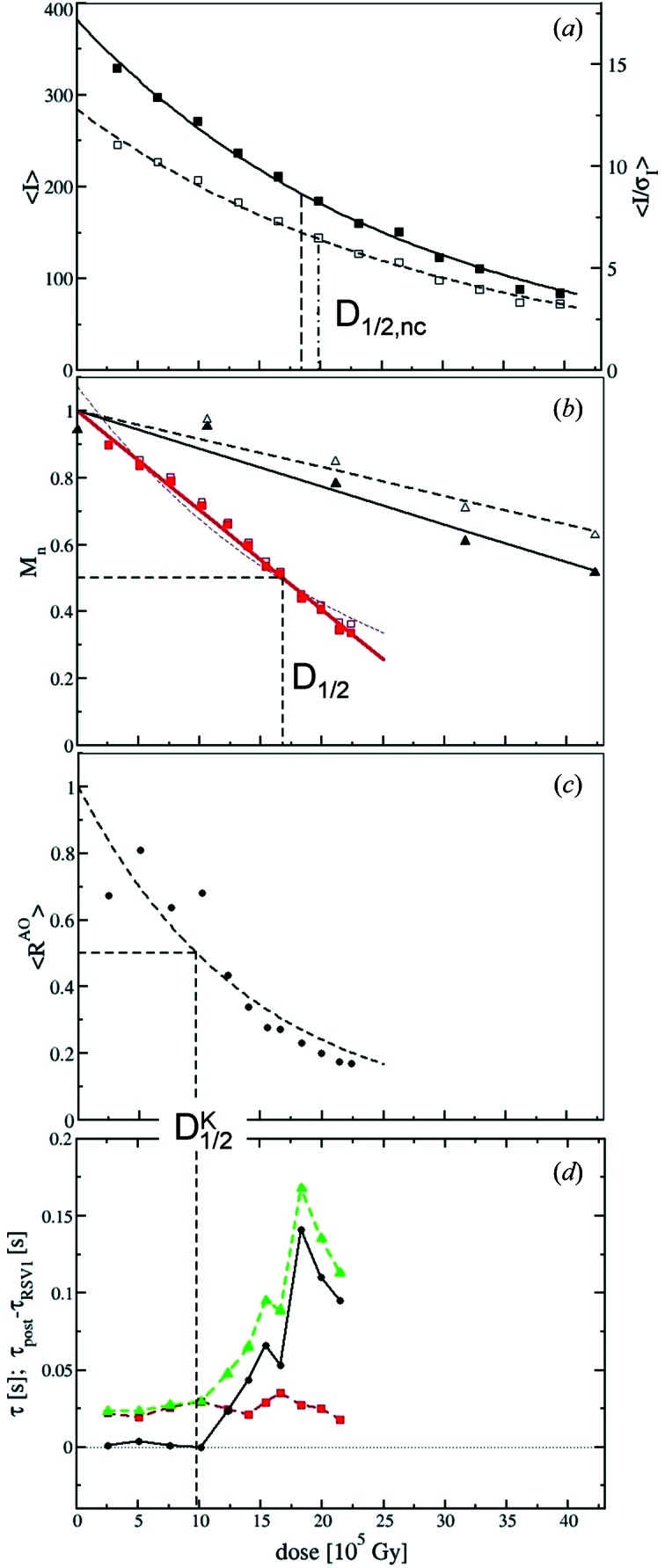
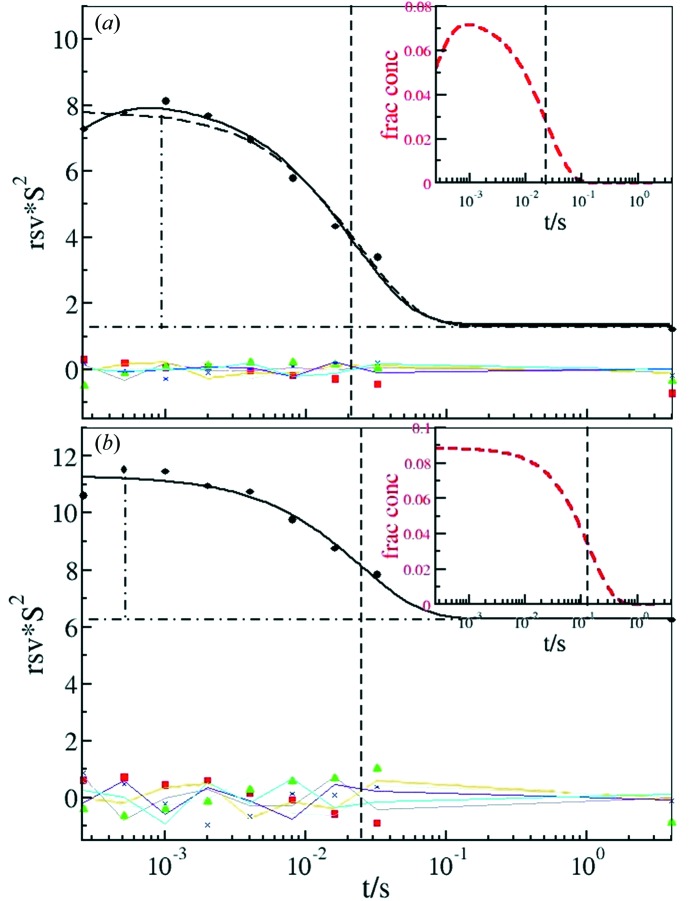
Protein X-ray structures are determined with ionizing radiation that damages the protein at high X-ray doses. As a result, diffraction patterns deteriorate with the increased absorbed dose. Several strategies such as sample freezing or scavenging of X-ray-generated free radicals are currently employed to minimize this damage. However, little is known about how the absorbed X-ray dose affects time-resolved Laue data collected at physiological temperatures where the protein is fully functional in the crystal, and how the kinetic analysis of such data depends on the absorbed dose. Here, direct evidence for the impact of radiation damage on the function of a protein is presented using time-resolved macromolecular crystallography. The effect of radiation damage on the kinetic analysis of time-resolved X-ray data is also explored.
蛋白质的 X 射线结构是用电离辐射来确定的,这种辐射会在高 X 射线剂量下损伤蛋白质。因此,随着吸收剂量的增加,衍射图案会恶化。目前,人们采用了几种策略,如样品冷冻或清除 X 射线产生的自由基,以尽量减少这种损伤。然而,对于吸收的 X 射线剂量如何影响在生理温度下收集的时间分辨劳埃数据,以及这种数据的动力学分析如何依赖于吸收剂量,人们知之甚少。在这里,使用时间分辨大分子晶体学直接证明了辐射损伤对蛋白质功能的影响。还探讨了辐射损伤对时间分辨 X 射线数据的动力学分析的影响。