• 文献检索
  • 文档翻译
  • 深度研究
  • 学术资讯
  • Suppr Zotero 插件Zotero 插件
  • 邀请有礼
  • 套餐&价格
  • 历史记录
应用&插件
Suppr Zotero 插件Zotero 插件浏览器插件Mac 客户端Windows 客户端微信小程序
定价
高级版会员购买积分包购买API积分包
服务
文献检索文档翻译深度研究API 文档MCP 服务
关于我们
关于 Suppr公司介绍联系我们用户协议隐私条款
关注我们

Suppr 超能文献

核心技术专利:CN118964589B侵权必究
粤ICP备2023148730 号-1Suppr @ 2026

文献检索

告别复杂PubMed语法,用中文像聊天一样搜索,搜遍4000万医学文献。AI智能推荐,让科研检索更轻松。

立即免费搜索

文件翻译

保留排版,准确专业,支持PDF/Word/PPT等文件格式,支持 12+语言互译。

免费翻译文档

深度研究

AI帮你快速写综述,25分钟生成高质量综述,智能提取关键信息,辅助科研写作。

立即免费体验

使用正电子发射断层扫描(PET)监测接受粒子治疗的患者:MIRANDA 研究。

Monitoring of patients treated with particle therapy using positron-emission-tomography (PET): the MIRANDA study.

机构信息

Department of Radiation Oncology, University Hospital of Heidelberg, Germany.

出版信息

BMC Cancer. 2012 Apr 3;12:133. doi: 10.1186/1471-2407-12-133.

DOI:10.1186/1471-2407-12-133
PMID:22471947
原文链接:https://pmc.ncbi.nlm.nih.gov/articles/PMC3350391/
Abstract

BACKGROUND

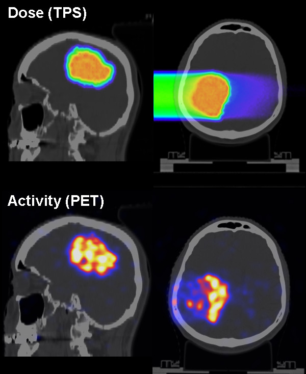
The purpose of this clinical study is to investigate the clinical feasibility and effectiveness of offline Positron-Emission-Tomography (PET) quality assurance for promoting the accuracy of proton and carbon ion beam therapy.

METHODS/DESIGN: A total of 240 patients will be recruited, evenly sampled among different analysis groups including tumors of the brain, skull base, head and neck region, upper gastrointestinal tract including the liver, lower gastrointestinal tract, prostate and pelvic region. From the comparison of the measured activity with the planned dose and its corresponding simulated activity distribution, conclusions on the delivered treatment will be inferred and, in case of significant deviations, correction strategies will be elaborated.

DISCUSSION

The investigated patients are expected to benefit from this study, since in case of detected deviations between planned and actual treatment delivery a proper intervention (e.g., correction) could be performed in a subsequent irradiation fraction. In this way, an overall better treatment could be achieved than without any in-vivo verification. Moreover, site-specific patient-population information on the precision of the ion range at HIT might enable improvement of the CT-range calibration curve as well as safe reduction of the treatment margins to promote enhanced treatment plan conformality and dose escalation for full clinical exploitation of the promises of ion beam therapy.

TRIAL REGISTRATION

NCT01528670.

摘要

背景

本临床研究旨在探讨离线正电子发射断层扫描(PET)质量保证在提高质子和碳离子束治疗准确性方面的临床可行性和有效性。

方法/设计:将招募 240 名患者,平均分配到包括脑肿瘤、颅底、头颈部、上胃肠道(包括肝脏)、下胃肠道、前列腺和骨盆区域在内的不同分析组中。通过将测量的活性与计划剂量及其相应的模拟活性分布进行比较,可以推断出对所提供治疗的结论,如果存在明显偏差,则制定纠正策略。

讨论

预期研究中的患者将从中受益,因为如果在计划和实际治疗输送之间检测到偏差,可以在下一次照射部分中进行适当的干预(例如纠正)。这样,就可以实现比没有任何体内验证更好的整体治疗效果。此外,HIT 处离子射程精度的特定部位患者人群信息可以改进 CT 射程校准曲线,并安全减少治疗边界,以促进治疗计划的一致性和剂量升级,从而充分利用离子束治疗的承诺。

试验注册

NCT01528670。

https://cdn.ncbi.nlm.nih.gov/pmc/blobs/1fef/3350391/a091887a2f97/1471-2407-12-133-1.jpg

相似文献

1
Monitoring of patients treated with particle therapy using positron-emission-tomography (PET): the MIRANDA study.使用正电子发射断层扫描(PET)监测接受粒子治疗的患者:MIRANDA 研究。
BMC Cancer. 2012 Apr 3;12:133. doi: 10.1186/1471-2407-12-133.
2
Systematic analysis of biological and physical limitations of proton beam range verification with offline PET/CT scans.使用离线PET/CT扫描对质子束射程验证的生物学和物理限制进行系统分析。
Phys Med Biol. 2009 Jul 21;54(14):4477-95. doi: 10.1088/0031-9155/54/14/008. Epub 2009 Jun 26.
3
In vivo verification of proton beam path by using post-treatment PET/CT imaging.使用治疗后PET/CT成像对质子束路径进行体内验证。
Med Phys. 2009 Sep;36(9):4136-46. doi: 10.1118/1.3193677.
4
Patient study of in vivo verification of beam delivery and range, using positron emission tomography and computed tomography imaging after proton therapy.质子治疗后使用正电子发射断层扫描和计算机断层扫描成像对束流传输和射程进行体内验证的患者研究。
Int J Radiat Oncol Biol Phys. 2007 Jul 1;68(3):920-34. doi: 10.1016/j.ijrobp.2007.01.063.
5
Accuracy of proton beam range verification using post-treatment positron emission tomography/computed tomography as function of treatment site.基于治疗部位的后处理正电子发射断层扫描/计算机断层扫描对质子束射程验证的准确性。
Int J Radiat Oncol Biol Phys. 2011 Jan 1;79(1):297-304. doi: 10.1016/j.ijrobp.2010.02.017. Epub 2010 Jun 18.
6
Comparison between in-beam and offline positron emission tomography imaging of proton and carbon ion therapeutic irradiation at synchrotron- and cyclotron-based facilities.基于同步加速器和回旋加速器的设施中质子和碳离子治疗性照射的在线与离线正电子发射断层扫描成像的比较。
Int J Radiat Oncol Biol Phys. 2008 Jul 1;71(3):945-56. doi: 10.1016/j.ijrobp.2008.02.033.
7
Implementation and initial clinical experience of offline PET/CT-based verification of scanned carbon ion treatment.基于离线 PET/CT 的扫描碳离子治疗验证的实施和初步临床经验。
Radiother Oncol. 2013 May;107(2):218-26. doi: 10.1016/j.radonc.2013.02.018. Epub 2013 May 4.
8
Development of dose delivery verification by PET imaging of photonuclear reactions following high energy photon therapy.通过高能光子治疗后光核反应的正电子发射断层显像(PET)成像进行剂量传递验证的研究进展。
Phys Med Biol. 2006 Nov 21;51(22):5769-83. doi: 10.1088/0031-9155/51/22/004. Epub 2006 Oct 19.
9
A filtering approach based on Gaussian-powerlaw convolutions for local PET verification of proton radiotherapy.一种基于高斯-幂律卷积的滤波方法用于质子放疗的局部正电子发射断层扫描(PET)验证
Phys Med Biol. 2006 Apr 21;51(8):1991-2009. doi: 10.1088/0031-9155/51/8/003. Epub 2006 Mar 30.
10
Automated evaluation of setup errors in carbon ion therapy using PET: feasibility study.基于正电子发射断层扫描的碳离子治疗摆位误差自动评估:可行性研究。
Med Phys. 2013 Dec;40(12):121718. doi: 10.1118/1.4829595.

引用本文的文献

1
Carbon ion radiotherapy with pencil beam scanning for hepatocellular carcinoma: Long-term outcomes from a phase I trial.碳离子放疗联合笔形束扫描治疗肝细胞癌:一项 I 期临床试验的长期结果。
Cancer Sci. 2023 Mar;114(3):976-983. doi: 10.1111/cas.15633. Epub 2022 Nov 15.
2
Technical Design Report for a Carbon-11 Treatment Facility.碳-11治疗设施技术设计报告
Front Med (Lausanne). 2022 Apr 25;8:697235. doi: 10.3389/fmed.2021.697235. eCollection 2021.
3
Monte Carlo investigation of the characteristics of radioactive beams for heavy ion therapy.

本文引用的文献

1
Implementation and initial clinical experience of offline PET/CT-based verification of scanned carbon ion treatment.基于离线 PET/CT 的扫描碳离子治疗验证的实施和初步临床经验。
Radiother Oncol. 2013 May;107(2):218-26. doi: 10.1016/j.radonc.2013.02.018. Epub 2013 May 4.
2
Implementation and workflow for PET monitoring of therapeutic ion irradiation: a comparison of in-beam, in-room, and off-line techniques.治疗性离子辐照的 PET 监测的实施和工作流程:在束、在室和离线技术的比较。
Phys Med Biol. 2011 Mar 7;56(5):1281-98. doi: 10.1088/0031-9155/56/5/004. Epub 2011 Feb 1.
3
Experimental verification of proton beam monitoring in a human body by use of activity image of positron-emitting nuclei generated by nuclear fragmentation reaction.
蒙特卡罗方法研究重离子治疗用放射性束的特性。
Sci Rep. 2019 Apr 25;9(1):6537. doi: 10.1038/s41598-019-43073-1.
4
MRI response of obturator internus muscle to carbon-ion dose in prostate cancer treatment.MRI 评估前列腺癌碳离子治疗中闭孔内肌的剂量反应。
J Radiat Res. 2019 May 1;60(3):335-341. doi: 10.1093/jrr/rry111.
5
Carbon ion beam radiotherapy for sinonasal malignant tumors invading skull base.碳离子束放射治疗侵犯颅底的鼻窦恶性肿瘤。
Case Rep Otolaryngol. 2014;2014:241856. doi: 10.1155/2014/241856. Epub 2014 Jun 9.
6
Proton therapy verification with PET imaging.正电子发射断层扫描成像技术在质子治疗中的验证。
Theranostics. 2013 Sep 19;3(10):731-40. doi: 10.7150/thno.5162.
利用核裂变反应产生的正电子发射核的活性图像对人体质子束监测进行实验验证。
Radiol Phys Technol. 2008 Jan;1(1):44-54. doi: 10.1007/s12194-007-0008-8. Epub 2007 Nov 27.
4
Randomized phase II study evaluating a carbon ion boost applied after combined radiochemotherapy with temozolomide versus a proton boost after radiochemotherapy with temozolomide in patients with primary glioblastoma: the CLEOPATRA trial.随机 II 期研究评估了在替莫唑胺联合放化疗后应用碳离子推量与在替莫唑胺放化疗后应用质子推量治疗原发性胶质母细胞瘤患者的疗效:CLEOPATRA 试验。
BMC Cancer. 2010 Sep 6;10:478. doi: 10.1186/1471-2407-10-478.
5
Radiological use of fast protons.快速质子的放射学应用。
Radiology. 1946 Nov;47(5):487-91. doi: 10.1148/47.5.487.
6
The development and clinical use of a beam ON-LINE PET system mounted on a rotating gantry port in proton therapy.一种安装在质子治疗旋转机架端口上的在线束 PET 系统的开发和临床应用。
Int J Radiat Oncol Biol Phys. 2010 Jan 1;76(1):277-86. doi: 10.1016/j.ijrobp.2009.05.065.
7
In vivo verification of proton beam path by using post-treatment PET/CT imaging.使用治疗后PET/CT成像对质子束路径进行体内验证。
Med Phys. 2009 Sep;36(9):4136-46. doi: 10.1118/1.3193677.
8
In-beam PET monitoring of mono-energetic (16)O and (12)C beams: experiments and FLUKA simulations for homogeneous targets.单能(16)O和(12)C束流的束内正电子发射断层扫描监测:均匀靶体的实验与FLUKA模拟
Phys Med Biol. 2009 Jul 7;54(13):3979-96. doi: 10.1088/0031-9155/54/13/003. Epub 2009 Jun 3.
9
Clinical CT-based calculations of dose and positron emitter distributions in proton therapy using the FLUKA Monte Carlo code.使用FLUKA蒙特卡罗代码基于临床CT进行质子治疗中剂量和正电子发射体分布的计算。
Phys Med Biol. 2007 Jun 21;52(12):3369-87. doi: 10.1088/0031-9155/52/12/004. Epub 2007 May 17.
10
Patient study of in vivo verification of beam delivery and range, using positron emission tomography and computed tomography imaging after proton therapy.质子治疗后使用正电子发射断层扫描和计算机断层扫描成像对束流传输和射程进行体内验证的患者研究。
Int J Radiat Oncol Biol Phys. 2007 Jul 1;68(3):920-34. doi: 10.1016/j.ijrobp.2007.01.063.