Department of Anatomy, Showa University School of Medicine, 15-8 Hatanodai, Shinagawa-ku, Tokyo 142-8555, Japan.
J Neuroinflammation. 2012 Apr 7;9:65. doi: 10.1186/1742-2094-9-65.
Microglia and macrophages (MG/MΦ) have a diverse range of functions depending on unique cytokine stimuli, and contribute to neural cell death, repair, and remodeling during central nervous system diseases. While IL-1 has been shown to exacerbate inflammation, it has also been recognized to enhance neuroregeneration. We determined the activating phenotype of MG/MΦ and the impact of IL-1 in an in vivo spinal cord injury (SCI) model of IL-1 knock-out (KO) mice. Moreover, we demonstrated the contribution of IL-1 to both the classical and alternative activation of MG in vitro using an adult MG primary culture.
SCI was induced by transection of the spinal cord between the T9 and T10 vertebra in wild-type and IL-1 KO mice. Locomotor activity was monitored and lesion size was determined for 14 days. TNFα and Ym1 levels were monitored to determine the MG/MΦ activating phenotype. Primary cultures of MG were produced from adult mice, and were exposed to IFNγ or IL-4 with and without IL-1β. Moreover, cultures were exposed to IL-4 and/or IL-13 in the presence and absence of IL-1β.
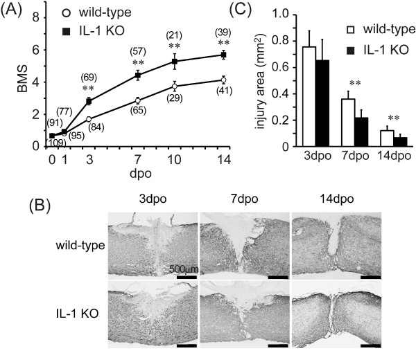
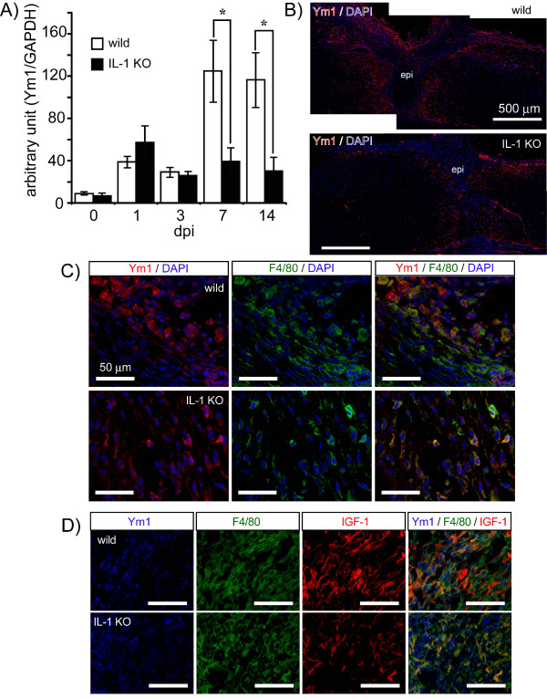
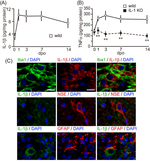
The locomotor activity and lesion area of IL-1 KO mice improved significantly after SCI compared with wild-type mice. TNFα production was significantly suppressed in IL-1 KO mice. Also, Ym1, an alternative activating MG/MΦ marker, did not increase in IL-1 KO mice, suggesting that IL-1 contributes to both the classical and alternative activation of MG/MΦ. We treated primary MG cultures with IFNγ or IL-4 in the presence and absence of IL-1β. Increased nitric oxide and TNFα was present in the culture media and increased inducible NO synthase was detected in cell suspensions following co-treatment with IFNγ and IL-1β. Expression of the alternative activation markers Ym1 and arginase-1 was increased after exposure to IL-4 and further increased after co-treatment with IL-4 and IL-1β. The phenotype was not observed after exposure of cells to IL-13.
We demonstrate here in in vivo experiments that IL-1 suppressed SCI in a process mediated by the reduction of inflammatory responses. Moreover, we suggest that IL-1 participates in both the classical and alternative activation of MG in in vivo and in vitro systems.
小胶质细胞和巨噬细胞(MG/MΦ)具有多种功能,取决于独特的细胞因子刺激,并有助于中枢神经系统疾病中的神经细胞死亡、修复和重塑。虽然 IL-1 已被证明会加剧炎症,但它也被认为可以增强神经再生。我们确定了 MG/MΦ 的激活表型,以及 IL-1 在 IL-1 敲除(KO)小鼠体内脊髓损伤(SCI)模型中的作用。此外,我们使用成人 MG 原代培养物证明了 IL-1 对 MG 经典和替代激活的贡献。
在野生型和 IL-1 KO 小鼠的 T9 和 T10 椎骨之间横断脊髓,诱导 SCI。监测运动活动并确定 14 天的损伤大小。监测 TNFα 和 Ym1 水平以确定 MG/MΦ 的激活表型。从成年小鼠中产生 MG 原代培养物,并在存在和不存在 IL-1β 的情况下暴露于 IFNγ 或 IL-4。此外,在存在和不存在 IL-1β 的情况下,培养物暴露于 IL-4 和/或 IL-13。
与野生型小鼠相比,IL-1 KO 小鼠的 SCI 后运动活动和损伤面积显著改善。IL-1 KO 小鼠中 TNFα 的产生明显受到抑制。此外,Ym1,一种替代激活的 MG/MΦ 标志物,在 IL-1 KO 小鼠中没有增加,这表明 IL-1 有助于 MG/MΦ 的经典和替代激活。我们用 IFNγ 或 IL-4 处理原代 MG 培养物,同时存在和不存在 IL-1β。在 IFNγ 和 IL-1β 共同处理后,培养基中存在增加的一氧化氮和 TNFα,细胞悬液中检测到诱导型一氧化氮合酶增加。在暴露于 IL-4 后,替代激活标志物 Ym1 和精氨酸酶-1 的表达增加,在与 IL-4 和 IL-1β 共同处理后进一步增加。在暴露于 IL-13 后,未观察到表型。
我们在体内实验中证明,IL-1 通过降低炎症反应来抑制 SCI。此外,我们表明 IL-1 参与了体内和体外系统中 MG 的经典和替代激活。