• 文献检索
  • 文档翻译
  • 深度研究
  • 学术资讯
  • Suppr Zotero 插件Zotero 插件
  • 邀请有礼
  • 套餐&价格
  • 历史记录
应用&插件
Suppr Zotero 插件Zotero 插件浏览器插件Mac 客户端Windows 客户端微信小程序
定价
高级版会员购买积分包购买API积分包
服务
文献检索文档翻译深度研究API 文档MCP 服务
关于我们
关于 Suppr公司介绍联系我们用户协议隐私条款
关注我们

Suppr 超能文献

核心技术专利:CN118964589B侵权必究
粤ICP备2023148730 号-1Suppr @ 2026

文献检索

告别复杂PubMed语法,用中文像聊天一样搜索,搜遍4000万医学文献。AI智能推荐,让科研检索更轻松。

立即免费搜索

文件翻译

保留排版,准确专业,支持PDF/Word/PPT等文件格式,支持 12+语言互译。

免费翻译文档

深度研究

AI帮你快速写综述,25分钟生成高质量综述,智能提取关键信息,辅助科研写作。

立即免费体验

使用ζ校准扫描隧道显微镜测定生物样品的厚度

Thickness determination of biological samples with a zeta-calibrated scanning tunneling microscope.

作者信息

Wang Z H, Hartmann T, Baumeister W, Guckenberger R

机构信息

Max-Planck-Institut für Biochemie, Abteilung Molekulare Strukturbiologie, Martinsried, Federal Republic of Germany.

出版信息

Proc Natl Acad Sci U S A. 1990 Dec;87(23):9343-7. doi: 10.1073/pnas.87.23.9343.

DOI:10.1073/pnas.87.23.9343
PMID:2251276
原文链接:https://pmc.ncbi.nlm.nih.gov/articles/PMC55161/
Abstract

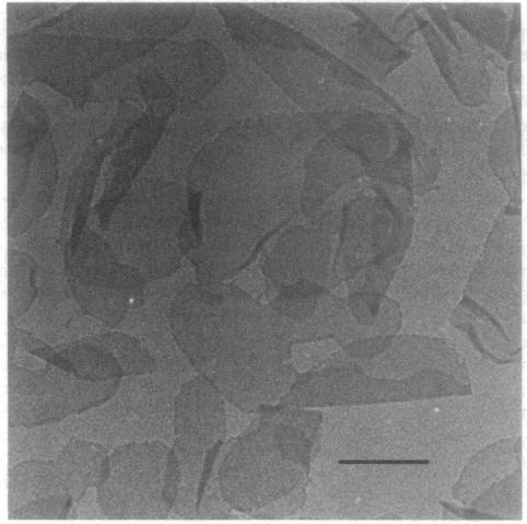
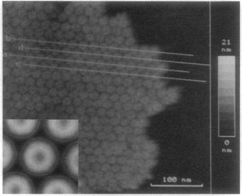
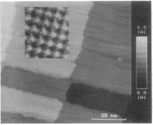
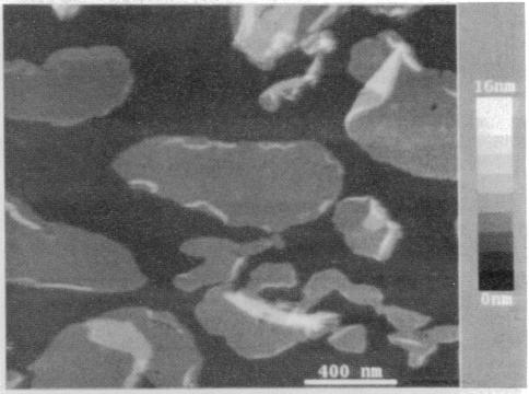
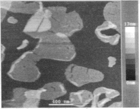
A single-tube scanning tunneling microscope has been zeta-calibrated by using atomic steps of crystalline gold and was used for measuring the thickness of two biological samples, metal-coated as well as uncoated. The hexagonal surface layer of the bacterium Deinococcus radiodurans with an open network-type structure shows thickness values that are strongly influenced by the substrate and the preparation method. In contrast, the thickness of the purple membrane of Halobacterium halobium with its densely packed less-corrugated structure exhibits very little variation in thickness in coated preparations and the values obtained are in good agreement with x-ray data.

摘要

一台单管扫描隧道显微镜已通过使用结晶金的原子台阶进行了ζ校准,并用于测量两个生物样品的厚度,这两个样品既有金属涂层的,也有未涂层的。具有开放网络型结构的耐辐射球菌的六边形表面层显示出的厚度值受到底物和制备方法的强烈影响。相比之下,具有紧密堆积且波纹较少结构的嗜盐菌紫膜在有涂层的制剂中厚度变化很小,所获得的值与X射线数据吻合良好。

https://cdn.ncbi.nlm.nih.gov/pmc/blobs/2d5c/55161/815473a0e1af/pnas01048-0283-a.jpg
https://cdn.ncbi.nlm.nih.gov/pmc/blobs/2d5c/55161/a522aac5b77f/pnas01048-0281-a.jpg
https://cdn.ncbi.nlm.nih.gov/pmc/blobs/2d5c/55161/401f28707f96/pnas01048-0281-b.jpg
https://cdn.ncbi.nlm.nih.gov/pmc/blobs/2d5c/55161/e8e898fb1394/pnas01048-0282-a.jpg
https://cdn.ncbi.nlm.nih.gov/pmc/blobs/2d5c/55161/5b4fdb1e218b/pnas01048-0282-b.jpg
https://cdn.ncbi.nlm.nih.gov/pmc/blobs/2d5c/55161/815473a0e1af/pnas01048-0283-a.jpg

相似文献

1
Thickness determination of biological samples with a zeta-calibrated scanning tunneling microscope.使用ζ校准扫描隧道显微镜测定生物样品的厚度
Proc Natl Acad Sci U S A. 1990 Dec;87(23):9343-7. doi: 10.1073/pnas.87.23.9343.
2
Measuring changes in membrane thickness by scanning tunneling microscopy.通过扫描隧道显微镜测量膜厚度的变化。
Biochim Biophys Acta. 1990 Apr 30;1023(3):325-34. doi: 10.1016/0005-2736(90)90123-6.
3
Purple membrane multilayers: detailed structure and photoelectric characteristics of bacteriorhodopsin.紫膜多层膜:细菌视紫红质的详细结构与光电特性
Biosystems. 1992;27(4):195-9. doi: 10.1016/0303-2647(92)90055-4.
4
Scanning tunneling microscopy of planar biomembranes.平面生物膜的扫描隧道显微镜检查
Ultramicroscopy. 1990 Aug;33(2):117-26. doi: 10.1016/0304-3991(90)90014-d.
5
Atomic force microscopy of purple membranes.紫膜的原子力显微镜观察
J Microsc. 1988 Dec;152(Pt 3):817-21. doi: 10.1111/j.1365-2818.1988.tb01454.x.
6
Masking generates contiguous segments of metal-coated and bare DNA for scanning tunneling microscope imaging.
Proc Natl Acad Sci U S A. 1993 Aug 15;90(16):7652-5. doi: 10.1073/pnas.90.16.7652.
7
Scanning probe microscopy. Probing the future.扫描探针显微镜。探索未来。
Science. 1997 Sep 19;277(5333):1845-7. doi: 10.1126/science.277.5333.1845.
8
Ionic channels in Langmuir-Blodgett films imaged by a scanning tunneling microscope.用扫描隧道显微镜成像的朗缪尔-布洛杰特膜中的离子通道。
Biophys J. 1991 Apr;59(4):889-93. doi: 10.1016/S0006-3495(91)82301-3.
9
Structural changes in native membrane proteins monitored at subnanometer resolution with the atomic force microscope: a review.利用原子力显微镜在亚纳米分辨率下监测天然膜蛋白的结构变化:综述
J Struct Biol. 1997 Jul;119(2):149-57. doi: 10.1006/jsbi.1997.3878.
10
Biological structures imaged in a hybrid scanning transmission electron microscope and scanning tunneling microscope.
Ultramicroscopy. 1991 Jun;35(3-4):255-64. doi: 10.1016/0304-3991(91)90077-j.

引用本文的文献

1
Tapping-mode atomic force microscopy produces faithful high-resolution images of protein surfaces.轻敲模式原子力显微镜可生成蛋白质表面逼真的高分辨率图像。
Biophys J. 1999 Aug;77(2):1150-8. doi: 10.1016/S0006-3495(99)76966-3.
2
High-resolution scanning tunneling microscopy of fully hydrated ripple-phase bilayers.完全水合波纹相双层膜的高分辨率扫描隧道显微镜观察
Biophys J. 1997 Feb;72(2 Pt 1):964-76. doi: 10.1016/s0006-3495(97)78731-9.
3
Vertical dimension of hydrated biological samples in tapping mode scanning force microscopy.轻敲模式扫描力显微镜下含水生物样品的垂直尺寸

本文引用的文献

1
Observation of atomic corrugation on Au(111) by scanning tunneling microscopy.通过扫描隧道显微镜对金(111)表面原子起伏的观测。
Phys Rev Lett. 1987 Dec 21;59(25):2879-2882. doi: 10.1103/PhysRevLett.59.2879.
2
The correlation averaging of a regularly arranged bacterial cell envelope protein.规则排列的细菌细胞包膜蛋白的相关性平均
J Microsc. 1982 Aug;127(Pt 2):127-38. doi: 10.1111/j.1365-2818.1982.tb00405.x.
3
The major cell envelope protein of Micrococcus radiodurans (R1). Structural and chemical characterization.耐辐射微球菌(R1)的主要细胞包膜蛋白。结构与化学特性
Biophys J. 1996 Mar;70(3):1514-20. doi: 10.1016/S0006-3495(96)79713-8.
4
Conformational change of the hexagonally packed intermediate layer of Deinococcus radiodurans monitored by atomic force microscopy.通过原子力显微镜监测耐辐射球菌六角形堆积中间层的构象变化。
J Bacteriol. 1996 Jun;178(11):3025-30. doi: 10.1128/jb.178.11.3025-3030.1996.
5
Covalent binding of biological samples to solid supports for scanning probe microscopy in buffer solution.生物样品在缓冲溶液中与固体支持物的共价结合用于扫描探针显微镜检查。
Biophys J. 1993 Dec;65(6):2437-46. doi: 10.1016/S0006-3495(93)81327-4.
6
Atomic force microscopy of three-dimensional membrane protein crystals. Ca-ATPase of sarcoplasmic reticulum.三维膜蛋白晶体的原子力显微镜观察。肌浆网钙ATP酶
Biophys J. 1992 Aug;63(2):303-8. doi: 10.1016/S0006-3495(92)81600-4.
7
The S-layer of Caulobacter crescentus: three-dimensional image reconstruction and structure analysis by electron microscopy.新月柄杆菌的S层:通过电子显微镜进行三维图像重建和结构分析
J Bacteriol. 1992 Oct;174(20):6527-38. doi: 10.1128/jb.174.20.6527-6538.1992.
Eur J Biochem. 1982 Jul;125(3):535-44. doi: 10.1111/j.1432-1033.1982.tb06715.x.
4
Rhodopsin-like protein from the purple membrane of Halobacterium halobium.来自嗜盐栖热菌紫膜的视紫红质样蛋白。
Nat New Biol. 1971 Sep 29;233(39):149-52. doi: 10.1038/newbio233149a0.
5
Isolation of the cell membrane of Halobacterium halobium and its fractionation into red and purple membrane.嗜盐菌细胞膜的分离及其分成红色膜和紫色膜的分级分离。
Methods Enzymol. 1974;31:667-78. doi: 10.1016/0076-6879(74)31072-5.
6
Three-dimensional structure of the regular surface layer (HPI layer) of Deinococcus radiodurans.耐辐射球菌规则表面层(HPI层)的三维结构。
J Mol Biol. 1986 Jan 20;187(2):241-50. doi: 10.1016/0022-2836(86)90231-7.
7
Electron-irradiation-induced flattening of negatively stained 2D protein crystals.电子辐照诱导的负染二维蛋白质晶体扁平化
Ultramicroscopy. 1987;21(4):313-9. doi: 10.1016/0304-3991(87)90029-5.
8
A scanning tunneling microscope (STM) for biological applications: design and performance.
Ultramicroscopy. 1988;25(2):111-21. doi: 10.1016/0304-3991(88)90218-5.
9
Scanning tunneling microscopy of recA-DNA complexes coated with a conducting film.对涂有导电膜的recA-DNA复合物进行扫描隧道显微镜观察。
Science. 1988 Apr 22;240(4851):514-6. doi: 10.1126/science.3358130.
10
Images of purple membrane at 2.8 A resolution obtained by cryo-electron microscopy.通过低温电子显微镜获得的分辨率为2.8埃的紫膜图像。
J Mol Biol. 1988 Aug 5;202(3):585-91. doi: 10.1016/0022-2836(88)90288-4.