Division of Bacteriology and Parasitology, Tulane National Primate Research Center, Covington, LA, USA.
J Neuroinflammation. 2012 Apr 23;9:72. doi: 10.1186/1742-2094-9-72.
Inflammation caused by the Lyme disease spirochete B. burgdorferi is an important factor in the pathogenesis of Lyme neuroborreliosis. Our central hypothesis is that B. burgdorferi can cause disease via the induction of inflammatory mediators such as cytokines and chemokines in glial and neuronal cells. Earlier we demonstrated that interaction of B. burgdorferi with brain parenchyma induces inflammatory mediators in glial cells as well as glial (oligodendrocyte) and neuronal apoptosis using ex vivo and in vivo models of experimentation.
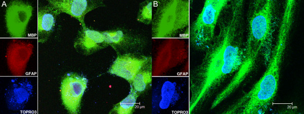
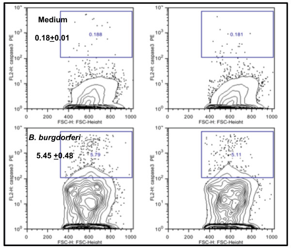
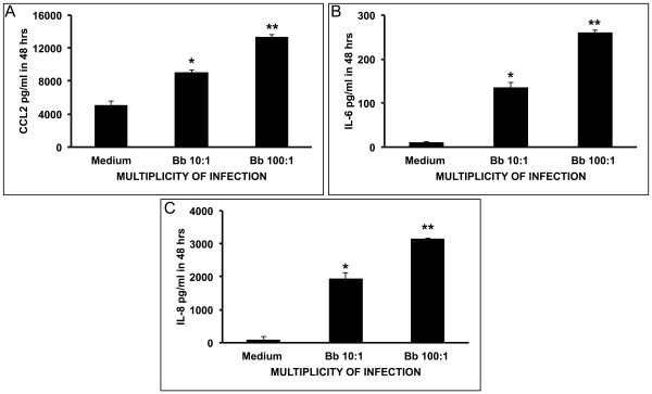
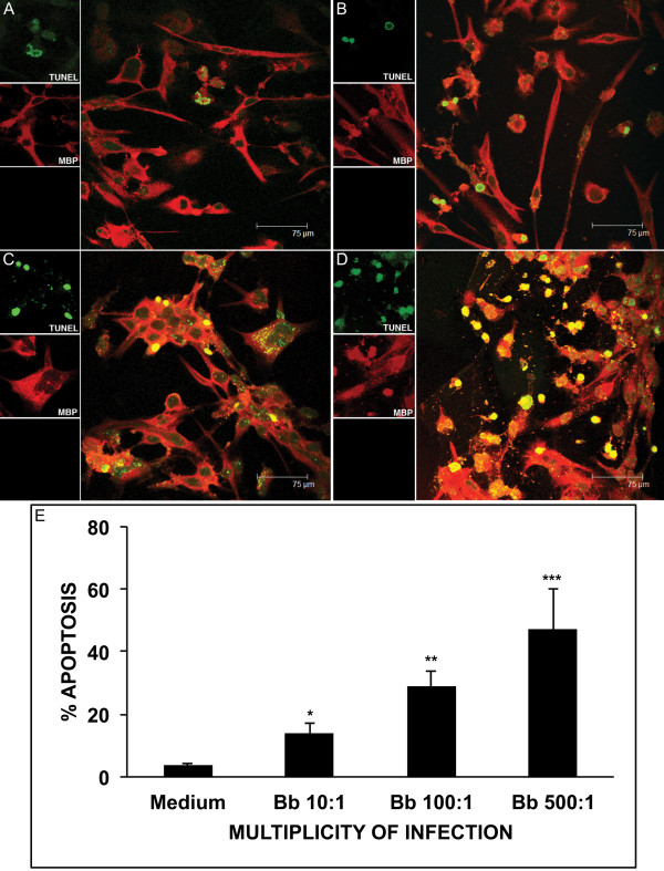
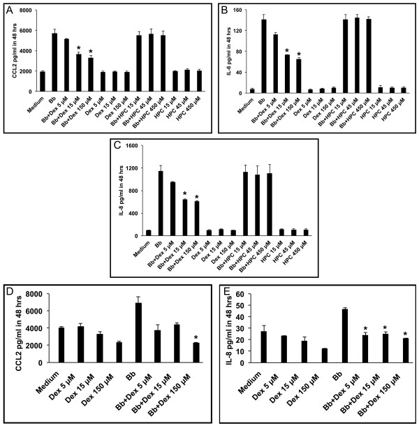
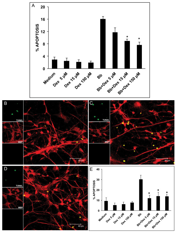
In this study we evaluated the ability of live B. burgdorferi to elicit inflammation in vitro in differentiated human MO3.13 oligodendrocytes and in differentiated primary human oligodendrocytes, by measuring the concentration of immune mediators in culture supernatants using Multiplex ELISA assays. Concomitant apoptosis was quantified in these cultures by the in situ terminal deoxynucleotidyl transferase mediated UTP nick end labeling (TUNEL) assay and by quantifying active caspase-3 by flow cytometry. The above phenomena were also evaluated after 48 h of stimulation with B. burgdorferi in the presence and absence of various concentrations of the anti-inflammatory drug dexamethasone.
B. burgdorferi induced enhanced levels of the cytokine IL-6 and the chemokines IL-8 and CCL2 in MO3.13 cells as compared to basal levels, and IL-8 and CCL2 in primary human oligodendrocytes, in a dose-dependent manner. These cultures also showed significantly elevated levels of apoptosis when compared with medium controls. Dexamethasone reduced both the levels of immune mediators and apoptosis, also in a manner that was dose dependent.
This finding supports our hypothesis that the inflammatory response elicited by the Lyme disease spirochete in glial cells contributes to neural cell damage. As oligodendrocytes are vital for the functioning and survival of neurons, the inflammation and subsequent apoptosis of oligodendrocytes induced by B. burgdorferi could contribute to the pathogenesis of Lyme neuroborreliosis.
莱姆病螺旋体伯氏疏螺旋体引起的炎症是莱姆神经Borreliosis 发病机制中的一个重要因素。我们的中心假设是,伯氏疏螺旋体可以通过诱导细胞因子和趋化因子等炎症介质在神经胶质细胞和神经元中引起疾病。我们之前的研究表明,伯氏疏螺旋体与脑实质的相互作用,使用离体和体内实验模型,诱导神经胶质细胞以及神经胶质(少突胶质细胞)和神经元凋亡中的炎症介质。
在这项研究中,我们通过使用多重 ELISA 测定法测量培养上清液中免疫介质的浓度,评估活伯氏疏螺旋体在体外分化的人 MO3.13 少突胶质细胞和分化的原代人少突胶质细胞中引起炎症的能力。在这些培养物中通过原位末端脱氧核苷酸转移酶介导的 UTP 缺口末端标记(TUNEL)测定和通过流式细胞术定量测定活性半胱天冬酶-3,同时定量凋亡。还在存在和不存在各种浓度的抗炎药物地塞米松的情况下,在 48 小时后评价上述现象。
与基础水平相比,伯氏疏螺旋体诱导 MO3.13 细胞中的细胞因子 IL-6 和趋化因子 IL-8 和 CCL2 以及原代人少突胶质细胞中的 IL-8 和 CCL2 水平增强,呈剂量依赖性。与培养基对照相比,这些培养物还显示出明显升高的凋亡水平。地塞米松降低了免疫介质和凋亡的水平,也是剂量依赖性的。
这一发现支持我们的假设,即莱姆病螺旋体在神经胶质细胞中引起的炎症反应导致神经细胞损伤。由于少突胶质细胞对神经元的功能和存活至关重要,因此伯氏疏螺旋体诱导的少突胶质细胞炎症和随后的凋亡可能有助于莱姆神经Borreliosis 的发病机制。