Paproki Anthony, Sirault Xavier, Berry Scott, Furbank Robert, Fripp Jurgen
The Australian e-Health Research Centre, CSIRO ICT Centre, Australia.
BMC Plant Biol. 2012 May 3;12:63. doi: 10.1186/1471-2229-12-63.
In recent years, imaging based, automated, non-invasive, and non-destructive high-throughput plant phenotyping platforms have become popular tools for plant biology, underpinning the field of plant phenomics. Such platforms acquire and record large amounts of raw data that must be accurately and robustly calibrated, reconstructed, and analysed, requiring the development of sophisticated image understanding and quantification algorithms. The raw data can be processed in different ways, and the past few years have seen the emergence of two main approaches: 2D image processing and 3D mesh processing algorithms. Direct image quantification methods (usually 2D) dominate the current literature due to comparative simplicity. However, 3D mesh analysis provides the tremendous potential to accurately estimate specific morphological features cross-sectionally and monitor them over-time.
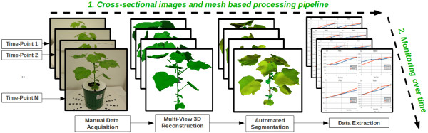
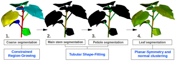
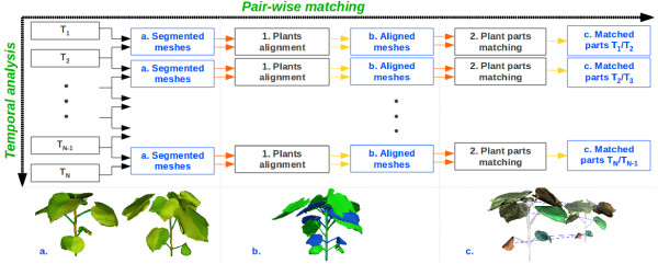
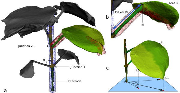
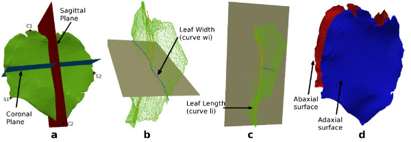
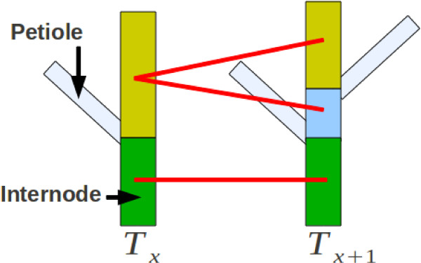
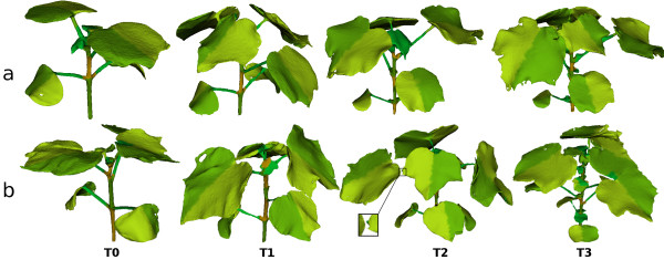
In this paper, we present a novel 3D mesh based technique developed for temporal high-throughput plant phenomics and perform initial tests for the analysis of Gossypium hirsutum vegetative growth. Based on plant meshes previously reconstructed from multi-view images, the methodology involves several stages, including morphological mesh segmentation, phenotypic parameters estimation, and plant organs tracking over time. The initial study focuses on presenting and validating the accuracy of the methodology on dicotyledons such as cotton but we believe the approach will be more broadly applicable. This study involved applying our technique to a set of six Gossypium hirsutum (cotton) plants studied over four time-points. Manual measurements, performed for each plant at every time-point, were used to assess the accuracy of our pipeline and quantify the error on the morphological parameters estimated.
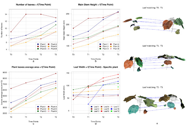
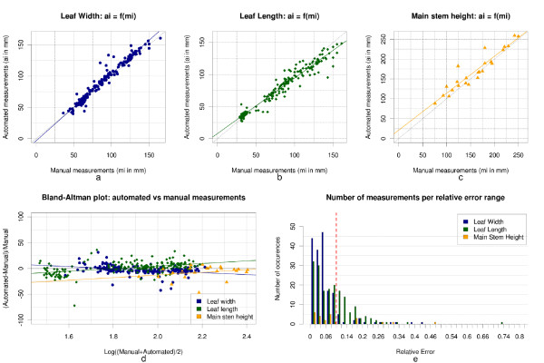
By directly comparing our automated mesh based quantitative data with manual measurements of individual stem height, leaf width and leaf length, we obtained the mean absolute errors of 9.34%, 5.75%, 8.78%, and correlation coefficients 0.88, 0.96, and 0.95 respectively. The temporal matching of leaves was accurate in 95% of the cases and the average execution time required to analyse a plant over four time-points was 4.9 minutes. The mesh processing based methodology is thus considered suitable for quantitative 4D monitoring of plant phenotypic features.
近年来,基于成像的、自动化的、非侵入性的和非破坏性的高通量植物表型分析平台已成为植物生物学领域的常用工具,为植物表型组学领域奠定了基础。此类平台获取并记录大量原始数据,这些数据必须经过准确且可靠的校准、重建和分析,这就需要开发复杂的图像理解和量化算法。原始数据可以通过不同方式进行处理,在过去几年中出现了两种主要方法:二维图像处理和三维网格处理算法。由于相对简单,直接图像量化方法(通常为二维)在当前文献中占主导地位。然而,三维网格分析具有巨大潜力,能够准确地从横截面估计特定形态特征并随时间进行监测。
在本文中,我们展示了一种为时间分辨高通量植物表型组学开发的基于三维网格的新技术,并对陆地棉营养生长分析进行了初步测试。基于先前从多视图图像重建的植物网格,该方法包括几个阶段,包括形态网格分割、表型参数估计以及植物器官随时间的跟踪。初步研究重点在于展示和验证该方法在双子叶植物如棉花上的准确性,但我们认为该方法将具有更广泛的适用性。本研究涉及将我们的技术应用于一组在四个时间点进行研究的六株陆地棉(棉花)植株。在每个时间点对每株植物进行的手动测量用于评估我们流程的准确性,并量化估计的形态参数上的误差。
通过将基于自动化网格的定量数据与单个茎高、叶宽和叶长经手动测量的数据直接比较,我们分别获得了9.34%、5.75%、8.78%的平均绝对误差以及0.88、0.96和0.95的相关系数。在95%的情况下,叶片的时间匹配是准确的,分析一株植物在四个时间点所需的平均执行时间为4.9分钟。因此,基于网格处理的方法被认为适用于植物表型特征的定量四维监测。