El Matri Leila, Chebil Ahmed, Kort Fedra, Bouraoui Rym, Largueche Leila, Mghaieth Fatma
Department of Ophthalmology, Hedi Rais Institute of Ophthalmology, Tunis, Tunisia.
J Ophthalmic Vis Res. 2010 Apr;5(2):127-9.
To describe optical coherence tomography (OCT) findings in a patient with Berlin's edema following blunt ocular trauma.
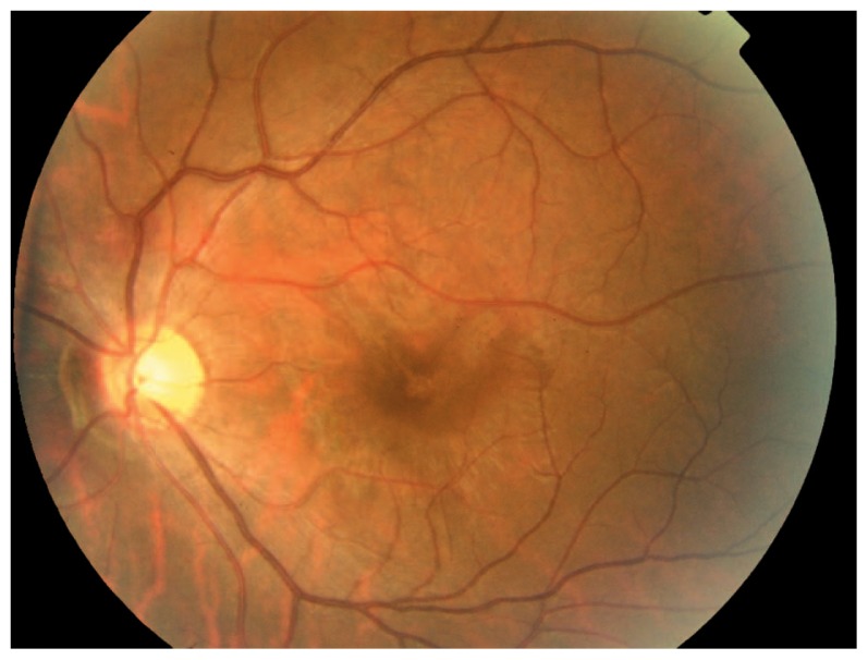
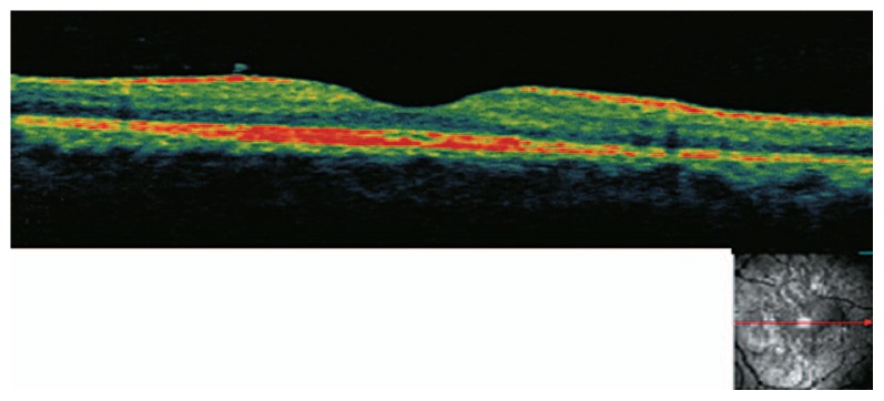
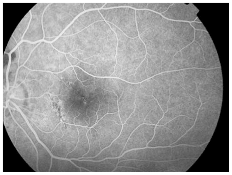
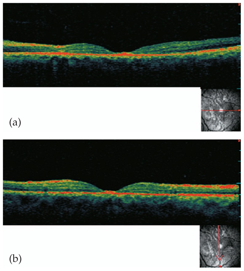
A 26-year-old man presented with acute loss of vision in his left eye following blunt trauma. He underwent a complete ophthalmologic examination and OCT. Fundus examination revealed abnormal yellow discoloration in the macula. OCT disclosed thickening of outer retinal structures and increased reflectivity in the area of photoreceptor outer segments with preservation of inner retinal architecture. Re-examination was conducted one month later at the time which OCT changes resolved leading to a surprisingly normal appearance.
OCT can be a useful tool in the diagnosis and follow-up of eyes with Berlin's edema and may reveal ultrastructural macular changes.
描述一名钝性眼外伤后发生柏林水肿患者的光学相干断层扫描(OCT)结果。
一名26岁男性在钝性外伤后左眼出现急性视力丧失。他接受了全面的眼科检查和OCT检查。眼底检查发现黄斑区有异常的黄色变色。OCT显示视网膜外层结构增厚,光感受器外段区域反射率增加,而视网膜内层结构保持完整。一个月后进行复查,此时OCT改变已消退,眼底外观出人意料地恢复正常。
OCT可作为诊断和随访柏林水肿眼的有用工具,并且可能揭示黄斑区的超微结构变化。