• 文献检索
  • 文档翻译
  • 深度研究
  • 学术资讯
  • Suppr Zotero 插件Zotero 插件
  • 邀请有礼
  • 套餐&价格
  • 历史记录
应用&插件
Suppr Zotero 插件Zotero 插件浏览器插件Mac 客户端Windows 客户端微信小程序
定价
高级版会员购买积分包购买API积分包
服务
文献检索文档翻译深度研究API 文档MCP 服务
关于我们
关于 Suppr公司介绍联系我们用户协议隐私条款
关注我们

Suppr 超能文献

核心技术专利:CN118964589B侵权必究
粤ICP备2023148730 号-1Suppr @ 2026

文献检索

告别复杂PubMed语法,用中文像聊天一样搜索,搜遍4000万医学文献。AI智能推荐,让科研检索更轻松。

立即免费搜索

文件翻译

保留排版,准确专业,支持PDF/Word/PPT等文件格式,支持 12+语言互译。

免费翻译文档

深度研究

AI帮你快速写综述,25分钟生成高质量综述,智能提取关键信息,辅助科研写作。

立即免费体验

通过对G蛋白偶联受体进行多序列和结构分析确定的相关突变网络内的一个最小配体结合口袋。

A minimal ligand binding pocket within a network of correlated mutations identified by multiple sequence and structural analysis of G protein coupled receptors.

作者信息

Moitra Subhodeep, Tirupula Kalyan C, Klein-Seetharaman Judith, Langmead Christopher James

机构信息

Computer Science Department, Carnegie Mellon University, Gates Hillman Center, 5000 Forbes Avenue, Pittsburgh, PA, USA.

Department of Structural Biology, University of Pittsburgh School of Medicine, Rm. 2051, Biomedical Science Tower 3, 3501 Fifth Avenue, Pittsburgh, PA, USA.

出版信息

BMC Biophys. 2012 Jun 29;5:13. doi: 10.1186/2046-1682-5-13.

DOI:10.1186/2046-1682-5-13
PMID:22748306
原文链接:https://pmc.ncbi.nlm.nih.gov/articles/PMC3478154/
Abstract

BACKGROUND

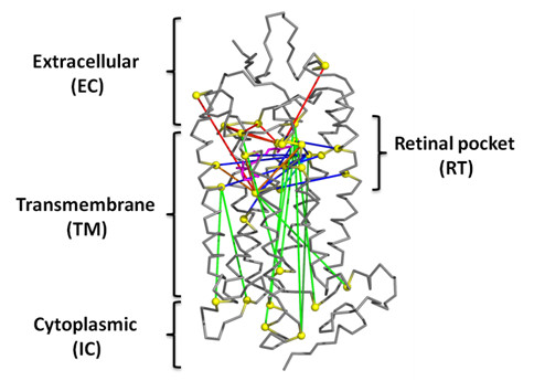
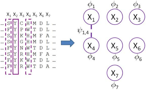
G protein coupled receptors (GPCRs) are seven helical transmembrane proteins that function as signal transducers. They bind ligands in their extracellular and transmembrane regions and activate cognate G proteins at their intracellular surface at the other side of the membrane. The relay of allosteric communication between the ligand binding site and the distant G protein binding site is poorly understood. In this study, GREMLIN 1, a recently developed method that identifies networks of co-evolving residues from multiple sequence alignments, was used to identify those that may be involved in communicating the activation signal across the membrane. The GREMLIN-predicted long-range interactions between amino acids were analyzed with respect to the seven GPCR structures that have been crystallized at the time this study was undertaken.

RESULTS

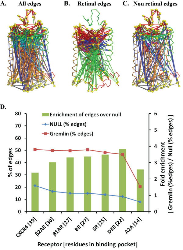
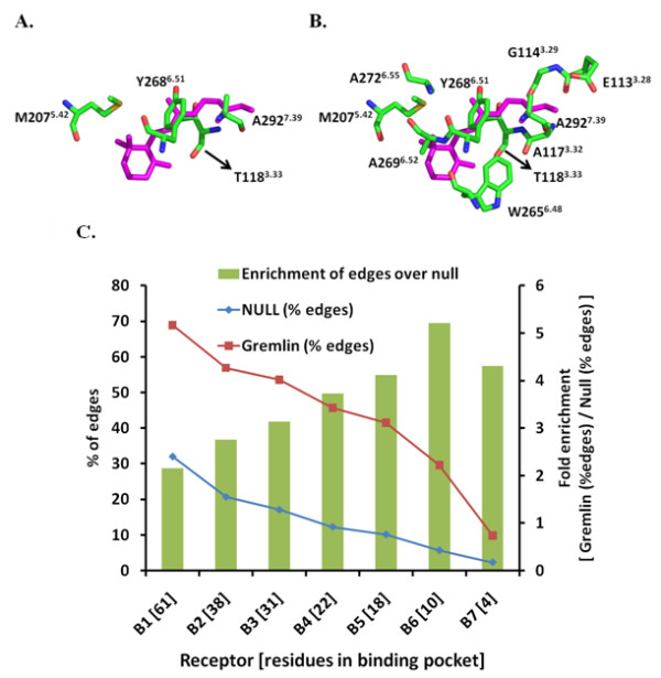
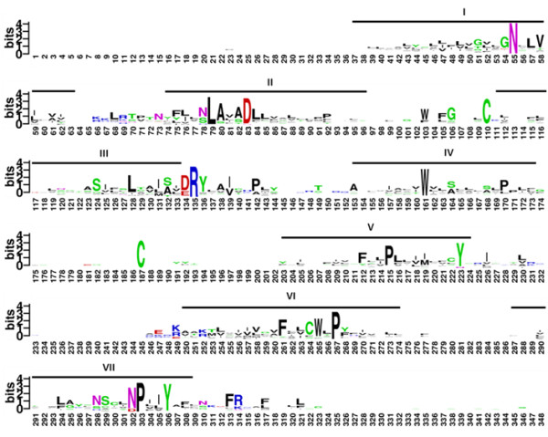
GREMLIN significantly enriches the edges containing residues that are part of the ligand binding pocket, when compared to a control distribution of edges drawn from a random graph. An analysis of these edges reveals a minimal GPCR binding pocket containing four residues (T1183.33, M2075.42, Y2686.51 and A2927.39). Additionally, of the ten residues predicted to have the most long-range interactions (A1173.32, A2726.55, E1133.28, H2115.46, S186EC2, A2927.39, E1223.37, G902.57, G1143.29 and M2075.42), nine are part of the ligand binding pocket.

CONCLUSIONS

We demonstrate the use of GREMLIN to reveal a network of statistically correlated and functionally important residues in class A GPCRs. GREMLIN identified that ligand binding pocket residues are extensively correlated with distal residues. An analysis of the GREMLIN edges across multiple structures suggests that there may be a minimal binding pocket common to the seven known GPCRs. Further, the activation of rhodopsin involves these long-range interactions between extracellular and intracellular domain residues mediated by the retinal domain.

摘要

背景

G蛋白偶联受体(GPCRs)是七螺旋跨膜蛋白,作为信号转导器发挥作用。它们在细胞外和跨膜区域结合配体,并在膜另一侧的细胞内表面激活同源G蛋白。配体结合位点与远处G蛋白结合位点之间的变构通讯传递机制尚不清楚。在本研究中,GREMLIN 1(一种最近开发的从多序列比对中识别共同进化残基网络的方法)被用于识别那些可能参与跨膜传递激活信号的残基。针对本研究开展时已结晶的七个GPCR结构,分析了GREMLIN预测的氨基酸之间的长程相互作用。

结果

与从随机图绘制的边的对照分布相比,GREMLIN显著富集了包含配体结合口袋一部分残基的边。对这些边的分析揭示了一个最小的GPCR结合口袋,包含四个残基(T1183.33、M2075.42、Y2686.51和A2927.39)。此外,在预测具有最长程相互作用的十个残基(A1173.32、A2726.55、E1133.28、H2115.46、S186EC2、A2927.39、E1223.37、G902.57、G1143.29和M2075.42)中,九个是配体结合口袋的一部分。

结论

我们展示了使用GREMLIN来揭示A类GPCR中具有统计学相关性和功能重要性的残基网络。GREMLIN确定配体结合口袋残基与远端残基广泛相关。对多个结构上GREMLIN边的分析表明,七个已知GPCR可能存在一个最小的共同结合口袋。此外,视紫红质的激活涉及由视黄醛结构域介导的细胞外和细胞内结构域残基之间的这些长程相互作用。

https://cdn.ncbi.nlm.nih.gov/pmc/blobs/a9dd/3478154/b0301b0308fe/2046-1682-5-13-7.jpg
https://cdn.ncbi.nlm.nih.gov/pmc/blobs/a9dd/3478154/a65da64dfac5/2046-1682-5-13-1.jpg
https://cdn.ncbi.nlm.nih.gov/pmc/blobs/a9dd/3478154/7a0b6de72005/2046-1682-5-13-2.jpg
https://cdn.ncbi.nlm.nih.gov/pmc/blobs/a9dd/3478154/2955cffbd564/2046-1682-5-13-3.jpg
https://cdn.ncbi.nlm.nih.gov/pmc/blobs/a9dd/3478154/7284aa5b3427/2046-1682-5-13-4.jpg
https://cdn.ncbi.nlm.nih.gov/pmc/blobs/a9dd/3478154/d07a4efe6377/2046-1682-5-13-5.jpg
https://cdn.ncbi.nlm.nih.gov/pmc/blobs/a9dd/3478154/0a3ceacda3cb/2046-1682-5-13-6.jpg
https://cdn.ncbi.nlm.nih.gov/pmc/blobs/a9dd/3478154/b0301b0308fe/2046-1682-5-13-7.jpg

相似文献

1
A minimal ligand binding pocket within a network of correlated mutations identified by multiple sequence and structural analysis of G protein coupled receptors.通过对G蛋白偶联受体进行多序列和结构分析确定的相关突变网络内的一个最小配体结合口袋。
BMC Biophys. 2012 Jun 29;5:13. doi: 10.1186/2046-1682-5-13.
2
Allosteric coupling and biased agonism in G protein-coupled receptors.变构偶联和 G 蛋白偶联受体的偏激动效应。
FEBS J. 2021 Apr;288(8):2513-2528. doi: 10.1111/febs.15783. Epub 2021 Mar 5.
3
7TM Domain Structure of Adhesion GPCRs.黏附G蛋白偶联受体的7次跨膜结构域
Handb Exp Pharmacol. 2016;234:43-66. doi: 10.1007/978-3-319-41523-9_3.
4
Evolutionary trace of G protein-coupled receptors reveals clusters of residues that determine global and class-specific functions.G蛋白偶联受体的进化踪迹揭示了决定整体和类别特异性功能的残基簇。
J Biol Chem. 2004 Feb 27;279(9):8126-32. doi: 10.1074/jbc.M312671200. Epub 2003 Dec 5.
5
Gonadotropin-Releasing Hormone (GnRH) Receptor Structure and GnRH Binding.促性腺激素释放激素(GnRH)受体结构与GnRH结合
Front Endocrinol (Lausanne). 2017 Oct 24;8:274. doi: 10.3389/fendo.2017.00274. eCollection 2017.
6
Agonist-induced conformational changes in bovine rhodopsin: insight into activation of G-protein-coupled receptors.激动剂诱导的牛视紫红质构象变化:深入了解G蛋白偶联受体的激活
J Mol Biol. 2008 Oct 3;382(2):539-55. doi: 10.1016/j.jmb.2008.06.084. Epub 2008 Jul 7.
7
Clusters of transmembrane residues are critical for human prostacyclin receptor activation.跨膜残基簇对人前列环素受体激活至关重要。
Biochemistry. 2004 Jul 20;43(28):8974-86. doi: 10.1021/bi0496788.
8
Conserved residues in RF-NH₂ receptor models identify predicted contact sites in ligand-receptor binding.RF-NH₂受体模型中的保守残基确定了配体-受体结合中的预测接触位点。
Peptides. 2014 Mar;53:278-85. doi: 10.1016/j.peptides.2013.06.009. Epub 2013 Jun 26.
9
Recognition of privileged structures by G-protein coupled receptors.G蛋白偶联受体对特殊结构的识别。
J Med Chem. 2004 Feb 12;47(4):888-99. doi: 10.1021/jm0309452.
10
Modeling of G-protein-coupled receptors: application to dopamine, adrenaline, serotonin, acetylcholine, and mammalian opsin receptors.G蛋白偶联受体的建模:应用于多巴胺、肾上腺素、血清素、乙酰胆碱和哺乳动物视蛋白受体。
J Med Chem. 1992 Sep 18;35(19):3448-62. doi: 10.1021/jm00097a002.

引用本文的文献

1
Transfer of knowledge from model organisms to evolutionarily distant non-model organisms: The coral Pocillopora damicornis membrane signaling receptome.从模式生物到进化上相距甚远的非模式生物的知识转移:珊瑚虫 Pocillopora damicornis 的膜信号受体组。
PLoS One. 2023 Feb 3;18(2):e0270965. doi: 10.1371/journal.pone.0270965. eCollection 2023.
2
Integrating structural and mutagenesis data to elucidate GPCR ligand binding.整合结构和诱变数据以阐明G蛋白偶联受体(GPCR)的配体结合情况。
Curr Opin Pharmacol. 2016 Oct;30:51-58. doi: 10.1016/j.coph.2016.07.003. Epub 2016 Jul 29.
3
Learning sequence determinants of protein:protein interaction specificity with sparse graphical models.

本文引用的文献

1
Learning generative models for protein fold families.学习蛋白质折叠家族的生成模型。
Proteins. 2011 Apr;79(4):1061-78. doi: 10.1002/prot.22934. Epub 2011 Jan 25.
2
Accounting for conformational entropy in predicting binding free energies of protein-protein interactions.在预测蛋白质-蛋白质相互作用的结合自由能时考虑构象熵。
Proteins. 2011 Feb;79(2):444-62. doi: 10.1002/prot.22894.
3
Structures of the CXCR4 chemokine GPCR with small-molecule and cyclic peptide antagonists.小分子和环肽拮抗剂与 CXCR4 趋化因子 GPCR 的结构。
利用稀疏图形模型学习蛋白质-蛋白质相互作用特异性的序列决定因素。
J Comput Biol. 2015 Jun;22(6):474-86. doi: 10.1089/cmb.2014.0289. Epub 2015 May 14.
4
Learning Sequence Determinants of Protein:protein Interaction Specificity with Sparse Graphical Models.利用稀疏图形模型研究蛋白质-蛋白质相互作用特异性的序列决定因素
Res Comput Mol Biol. 2014;8394:129-143. doi: 10.1007/978-3-319-05269-4_10.
5
Atypical signaling and functional desensitization response of MAS receptor to peptide ligands.MAS受体对肽配体的非典型信号传导和功能脱敏反应。
PLoS One. 2014 Jul 28;9(7):e103520. doi: 10.1371/journal.pone.0103520. eCollection 2014.
Science. 2010 Nov 19;330(6007):1066-71. doi: 10.1126/science.1194396. Epub 2010 Oct 7.
4
The Pfam protein families database.Pfam 蛋白质家族数据库。
Nucleic Acids Res. 2010 Jan;38(Database issue):D211-22. doi: 10.1093/nar/gkp985. Epub 2009 Nov 17.
5
Multiple switches in G protein-coupled receptor activation.G蛋白偶联受体激活中的多种开关机制
Trends Pharmacol Sci. 2009 Sep;30(9):494-502. doi: 10.1016/j.tips.2009.06.003. Epub 2009 Sep 3.
6
Helix movement is coupled to displacement of the second extracellular loop in rhodopsin activation.在视紫红质激活过程中,螺旋运动与第二个细胞外环的位移相关联。
Nat Struct Mol Biol. 2009 Feb;16(2):168-75. doi: 10.1038/nsmb.1549. Epub 2009 Feb 1.
7
Crystal structure of opsin in its G-protein-interacting conformation.视蛋白处于与G蛋白相互作用构象时的晶体结构。
Nature. 2008 Sep 25;455(7212):497-502. doi: 10.1038/nature07330.
8
Crystal structure of the ligand-free G-protein-coupled receptor opsin.无配体G蛋白偶联受体视蛋白的晶体结构。
Nature. 2008 Jul 10;454(7201):183-7. doi: 10.1038/nature07063. Epub 2008 Jun 18.
9
Protein structure modeling with MODELLER.使用MODELLER进行蛋白质结构建模。
Methods Mol Biol. 2008;426:145-59. doi: 10.1007/978-1-60327-058-8_8.
10
Graphical models of residue coupling in protein families.蛋白质家族中残基偶联的图形模型。
IEEE/ACM Trans Comput Biol Bioinform. 2008 Apr-Jun;5(2):183-97. doi: 10.1109/TCBB.2007.70225.