• 文献检索
  • 文档翻译
  • 深度研究
  • 学术资讯
  • Suppr Zotero 插件Zotero 插件
  • 邀请有礼
  • 套餐&价格
  • 历史记录
应用&插件
Suppr Zotero 插件Zotero 插件浏览器插件Mac 客户端Windows 客户端微信小程序
定价
高级版会员购买积分包购买API积分包
服务
文献检索文档翻译深度研究API 文档MCP 服务
关于我们
关于 Suppr公司介绍联系我们用户协议隐私条款
关注我们

Suppr 超能文献

核心技术专利:CN118964589B侵权必究
粤ICP备2023148730 号-1Suppr @ 2026

文献检索

告别复杂PubMed语法,用中文像聊天一样搜索,搜遍4000万医学文献。AI智能推荐,让科研检索更轻松。

立即免费搜索

文件翻译

保留排版,准确专业,支持PDF/Word/PPT等文件格式,支持 12+语言互译。

免费翻译文档

深度研究

AI帮你快速写综述,25分钟生成高质量综述,智能提取关键信息,辅助科研写作。

立即免费体验

常规胸部X光检查:在超级机器时代,对评估左心室大小和功能仍有价值吗?

Routine Chest X-ray: Still Valuable for the Assessment of Left Ventricular Size and Function in the Era of Super Machines?

作者信息

Morales Maria-Aurora, Prediletto Renato, Rossi Giuseppe, Catapano Giosuè, Lombardi Massimo, Rovai Daniele

机构信息

CNR, Clinical Physiology Institute, Pisa, Italy.

出版信息

J Clin Imaging Sci. 2012;2:25. doi: 10.4103/2156-7514.96540. Epub 2012 May 23.

DOI:10.4103/2156-7514.96540
PMID:22754739
原文链接:https://pmc.ncbi.nlm.nih.gov/articles/PMC3385501/
Abstract

OBJECTIVES

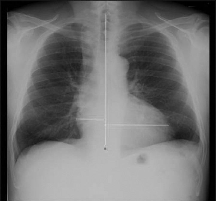
The development of technologically advanced, expensive techniques has progressively reduced the value of chest X-ray in clinical practice for the assessment of left ventricular (LV) dilatation and dysfunction. Although controversial data are reported on the role of this widely available technique in cardiac assessment, it is known that the cardio-thoracic ratio is predictive of risk of progression in the NYHA Class, hospitalization, and outcome in patients with LV dysfunction. This study aimed to evaluate the reliability of the transverse diameter of heart shadow [TDH] by chest X-ray for detecting LV dilatation and dysfunction as compared to Magnetic Resonance Imaging (MRI) performed for different clinical reasons.

MATERIALS AND METHODS

In 101 patients, TDH was measured in digital chest X-ray and LV volumes and ejection fraction (EF) by MRI, both exams performed within 2 days.

RESULTS

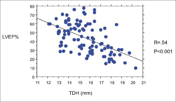
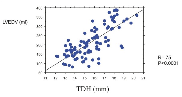
A direct correlation between TDH and end-diastolic volumes (r = .75, P<0.0001) was reported. TDH cut-off values of 14.5 mm in females identified LV end-diastolic volumes >150 mL (sensitivity: 82%, specificity: 69%); in males a cut-off value of 15.5 mm identified LV end-diastolic volumes >210 mL (sensitivity: 84%; specificity: 72%). A negative relation was found between TDH and LVEF (r = -.54, P<0.0001). The above cut-off values of TDH discriminated patients with LV systolic dysfunction - LVEF <35% (sensitivity and specificity: 67% and 57% in females; 76% and 59% in males, respectively).

CONCLUSIONS

Chest X-ray may still be considered a reliable technique in predicting LV dilatation by the accurate measurement of TDH as compared to cardiac MRI. Technologically advanced, expensive, and less available imaging techniques should be performed on the basis of sound clinical requests.

摘要

目的

技术先进、价格昂贵的技术发展已逐渐降低了胸部X线在临床实践中评估左心室(LV)扩张和功能障碍的价值。尽管关于这一广泛应用的技术在心脏评估中的作用存在有争议的数据,但已知心胸比率可预测纽约心脏协会(NYHA)心功能分级进展、住院情况及LV功能障碍患者的预后。本研究旨在评估胸部X线测量心脏阴影横径(TDH)检测LV扩张和功能障碍的可靠性,并与因不同临床原因进行的磁共振成像(MRI)相比较。

材料与方法

对101例患者进行了数字胸部X线TDH测量及MRI测量LV容积和射血分数(EF),两项检查均在2天内完成。

结果

报告TDH与舒张末期容积之间存在直接相关性(r = 0.75,P<0.0001)。女性TDH临界值为14.5 mm时,可识别舒张末期LV容积>150 mL(敏感性:82%,特异性:69%);男性临界值为15.5 mm时,可识别舒张末期LV容积>210 mL(敏感性:84%;特异性:72%)。TDH与左室射血分数呈负相关(r = -0.54,P<0.0001)。上述TDH临界值可区分LV收缩功能障碍(左室射血分数<35%)患者——女性的敏感性和特异性分别为67%和57%;男性分别为76%和59%。

结论

与心脏MRI相比,通过准确测量TDH,胸部X线在预测LV扩张方面仍可被视为一种可靠的技术。技术先进、价格昂贵且应用较少的成像技术应根据合理的临床需求进行。

https://cdn.ncbi.nlm.nih.gov/pmc/blobs/9b1a/3385501/9243675c01a3/JCIS-2-25-g004.jpg
https://cdn.ncbi.nlm.nih.gov/pmc/blobs/9b1a/3385501/eeb4477e9df4/JCIS-2-25-g002.jpg
https://cdn.ncbi.nlm.nih.gov/pmc/blobs/9b1a/3385501/c9bae782bc70/JCIS-2-25-g003.jpg
https://cdn.ncbi.nlm.nih.gov/pmc/blobs/9b1a/3385501/9243675c01a3/JCIS-2-25-g004.jpg
https://cdn.ncbi.nlm.nih.gov/pmc/blobs/9b1a/3385501/eeb4477e9df4/JCIS-2-25-g002.jpg
https://cdn.ncbi.nlm.nih.gov/pmc/blobs/9b1a/3385501/c9bae782bc70/JCIS-2-25-g003.jpg
https://cdn.ncbi.nlm.nih.gov/pmc/blobs/9b1a/3385501/9243675c01a3/JCIS-2-25-g004.jpg

相似文献

1
Routine Chest X-ray: Still Valuable for the Assessment of Left Ventricular Size and Function in the Era of Super Machines?常规胸部X光检查:在超级机器时代,对评估左心室大小和功能仍有价值吗?
J Clin Imaging Sci. 2012;2:25. doi: 10.4103/2156-7514.96540. Epub 2012 May 23.
2
Positron emission tomography for the assessment of myocardial viability: an evidence-based analysis.用于评估心肌活力的正电子发射断层扫描:一项基于证据的分析。
Ont Health Technol Assess Ser. 2005;5(16):1-167. Epub 2005 Oct 1.
3
Comparative value of ECG-gated blood pool SPET and ECG-gated myocardial perfusion SPET in the assessment of global systolic left ventricular function.心电图门控心血池单光子发射计算机断层显像(SPET)与心电图门控心肌灌注单光子发射计算机断层显像在评估左心室整体收缩功能中的比较价值
Eur J Nucl Med Mol Imaging. 2003 Jun;30(6):859-67. doi: 10.1007/s00259-003-1121-5. Epub 2003 Apr 4.
4
Quantitative Assessment of Right Ventricular Volumes and Ejection Fraction in Patients with Left Ventricular Systolic Dysfunction by Real Time Three-Dimensional Echocardiography versus Cardiac Magnetic Resonance Imaging.实时三维超声心动图与心脏磁共振成像对左心室收缩功能不全患者右心室容积和射血分数的定量评估
Echocardiography. 2015 May;32(5):805-12. doi: 10.1111/echo.12715. Epub 2014 Aug 11.
5
Comparison of left ventricular volumes and ejection fraction by monoplane cineventriculography, unenhanced echocardiography and cardiac magnetic resonance imaging.单平面电影心室造影、未增强超声心动图和心脏磁共振成像左心室容量和射血分数的比较。
Int J Cardiovasc Imaging. 2012 Jun;28(5):1003-10. doi: 10.1007/s10554-011-9924-0. Epub 2011 Jul 27.
6
Comparison Between 3D Echocardiography and Cardiac Magnetic Resonance Imaging (CMRI) in the Measurement of Left Ventricular Volumes and Ejection Fraction.三维超声心动图与心脏磁共振成像(CMRI)在测量左心室容积和射血分数方面的比较
Curr Med Imaging Rev. 2019;15(7):654-660. doi: 10.2174/1573405614666180815115756.
7
Real time three-dimensional echocardiographic assessment of left ventricular function in heart failure patients: underestimation of left ventricular volume increases with the degree of dilatation.心力衰竭患者左心室功能的实时三维超声心动图评估:左心室容积低估随扩张程度增加。
Echocardiography. 2012 Sep;29(8):970-7. doi: 10.1111/j.1540-8175.2012.01707.x. Epub 2012 May 8.
8
The heart as a spring, the measurement of myocardial bounce to assess left ventricular function on cardiac MR.心脏犹如弹簧,通过测量心肌回弹来评估心脏磁共振左心室功能。
Int J Cardiovasc Imaging. 2021 May;37(5):1699-1707. doi: 10.1007/s10554-020-02129-4. Epub 2021 Feb 23.
9
Accuracy of three-dimensional echocardiography with unrestricted selection of imaging planes for measurement of left ventricular volumes and ejection fraction.不受成像平面选择限制的三维超声心动图测量左心室容积和射血分数的准确性。
Am Heart J. 2000 Sep;140(3):469-75. doi: 10.1067/mhj.2000.108513.
10
[Left ventricular volume and ejection fraction by the axius auto ejection fraction method: comparison with manual trace method and visual assessment of ejection fraction].[采用Axius自动射血分数法测量左心室容积和射血分数:与手动描记法及射血分数视觉评估法的比较]
J Cardiol. 2007 Mar;49(3):125-34.

引用本文的文献

1
Novel Indexes in the Assessment of Cardiac Enlargement Using Chest Radiography: A New Look at an Old Problem.胸部X线摄影评估心脏扩大的新指标:重新审视一个老问题。
J Clin Med. 2025 Feb 1;14(3):942. doi: 10.3390/jcm14030942.
2
Cardiothoracic ratio and left ventricular ejection fraction relationship: A meta-analysis study.心胸比与左心室射血分数的关系:一项荟萃分析研究。
Saudi Med J. 2023 Jun;44(6):529-536. doi: 10.15537/smj.2023.44.6.20230027.
3
Clinical Data, Chest Radiograph and Electrocardiography in the Screening for Left Ventricular Hypertrophy: The CARE Score.

本文引用的文献

1
Evaluation of left ventricular volumes and ejection fraction by gated SPECT and cardiac MRI in patients with dilated cardiomyopathy.门控单光子发射计算机断层扫描(gated SPECT)和心脏磁共振成像(MRI)评估扩张型心肌病患者左心室容积和射血分数
Eur J Nucl Med Mol Imaging. 2009 Oct;36(10):1611-21. doi: 10.1007/s00259-009-1136-7. Epub 2009 Apr 18.
2
Non-invasive investigations of the right heart: how and why?右心的非侵入性检查:方法及原因?
Arch Cardiovasc Dis. 2009 Mar;102(3):219-32. doi: 10.1016/j.acvd.2008.12.010. Epub 2009 Mar 19.
3
Accuracy of chest radiographs in the emergency diagnosis of heart failure.
用于筛查左心室肥厚的临床数据、胸部X光片和心电图:CARE评分
J Clin Med. 2022 Jun 22;11(13):3585. doi: 10.3390/jcm11133585.
4
Gender and Age Differences in Cardiac Size Parameters of Ghanaian Adults: Can One Parameter Fit All? Part Two.加纳成年人心脏大小参数的性别和年龄差异:是否有一种参数适用于所有人?第二部分。
Ethiop J Health Sci. 2021 May;31(3):561-572. doi: 10.4314/ejhs.v31i3.13.
5
Revisiting anatomical variants on screening chest radiographs in Indian adolescents: A cross sectional observational pilot study.重新审视印度青少年胸部X光筛查中的解剖变异:一项横断面观察性试点研究。
Med J Armed Forces India. 2018 Oct;74(4):337-345. doi: 10.1016/j.mjafi.2017.07.010. Epub 2017 Aug 30.
6
Electrochemical Aptamer-Based Biosensors for the Detection of Cardiac Biomarkers.用于检测心脏生物标志物的基于电化学适配体的生物传感器。
ACS Omega. 2018 Sep 30;3(9):12010-12018. doi: 10.1021/acsomega.8b01558. Epub 2018 Sep 26.
7
Radiographic determination of cardiomegaly using cardiothoracic ratio and transverse cardiac diameter: can one size fit all? Part one.使用心胸比率和心脏横径进行心脏扩大的影像学判定:一种方法能适用于所有情况吗?第一部分。
Pan Afr Med J. 2017 Jul 14;27:201. doi: 10.11604/pamj.2017.27.201.12017. eCollection 2017.
8
Cardio-Thoracic Ratio Is Stable, Reproducible and Has Potential as a Screening Tool for HIV-1 Related Cardiac Disorders in Resource Poor Settings.心胸比率稳定、可重现,且有可能成为资源匮乏环境下 HIV-1 相关心脏疾病的筛查工具。
PLoS One. 2016 Oct 4;11(10):e0163490. doi: 10.1371/journal.pone.0163490. eCollection 2016.
胸部X光片在心力衰竭急诊诊断中的准确性。
Eur Radiol. 2008 Aug;18(8):1644-52. doi: 10.1007/s00330-008-0930-0. Epub 2008 Mar 28.
4
Classic imaging signs of congenital cardiovascular abnormalities.先天性心血管异常的典型影像学表现。
Radiographics. 2007 Sep-Oct;27(5):1323-34. doi: 10.1148/rg.275065148.
5
MRI of left ventricular function.左心室功能的磁共振成像
J Nucl Cardiol. 2007 Sep-Oct;14(5):729-44. doi: 10.1016/j.nuclcard.2007.07.006.
6
Imaging patients with heart failure: expectations of the clinician.
Heart Fail Clin. 2006 Apr;2(2):107-15. doi: 10.1016/j.hfc.2006.05.003.
7
[Imaging of pulmonary arterial hypertension].[肺动脉高压的影像学检查]
Rev Mal Respir. 2007 Feb;24(2):155-69.
8
Cardiovascular magnetic resonance imaging for the assessment of right heart involvement in cardiac and pulmonary disease.心血管磁共振成像用于评估心脏和肺部疾病中右心受累情况。
Heart Lung Circ. 2006 Dec;15(6):362-70. doi: 10.1016/j.hlc.2006.08.003. Epub 2006 Oct 11.
9
Diagnosis of heart failure in primary care.基层医疗中的心衰诊断。
Heart Fail Rev. 2006 Jun;11(2):95-107. doi: 10.1007/s10741-006-9481-0.
10
Radiographic measures of intravascular volume status: the role of vascular pedicle width.
Curr Opin Crit Care. 2006 Jun;12(3):255-62. doi: 10.1097/01.ccx.0000224871.31947.8d.