Department of Physics, University of California, Berkeley, CA 94720, USA.
Science. 2012 Jul 13;337(6091):236-9. doi: 10.1126/science.1222981.
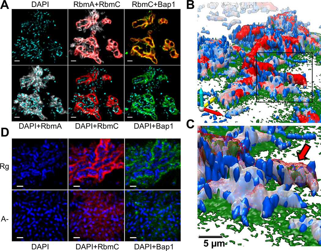
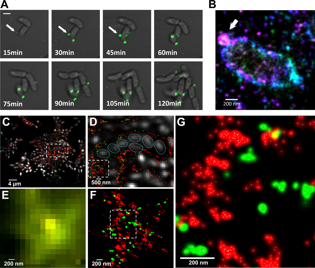
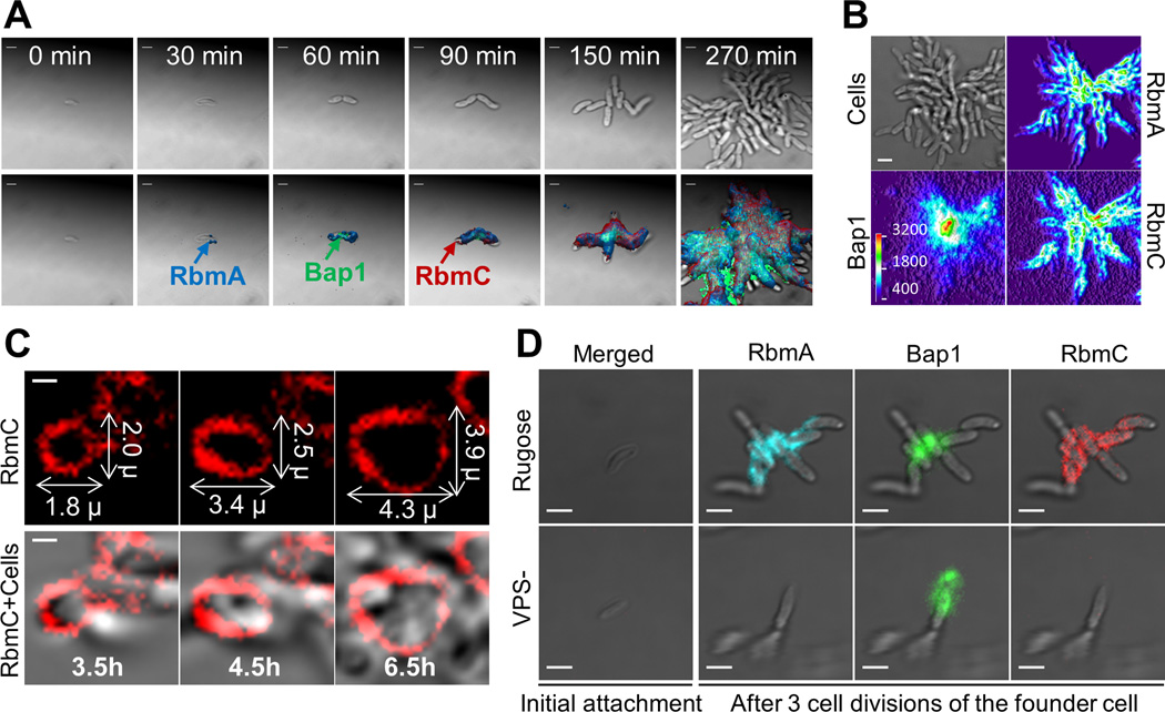
In their natural environment, microbes organize into communities held together by an extracellular matrix composed of polysaccharides and proteins. We developed an in vivo labeling strategy to allow the extracellular matrix of developing biofilms to be visualized with conventional and superresolution light microscopy. Vibrio cholerae biofilms displayed three distinct levels of spatial organization: cells, clusters of cells, and collections of clusters. Multiresolution imaging of living V. cholerae biofilms revealed the complementary architectural roles of the four essential matrix constituents: RbmA provided cell-cell adhesion; Bap1 allowed the developing biofilm to adhere to surfaces; and heterogeneous mixtures of Vibrio polysaccharide, RbmC, and Bap1 formed dynamic, flexible, and ordered envelopes that encased the cell clusters.
在自然环境中,微生物通过由多糖和蛋白质组成的细胞外基质聚集形成群落。我们开发了一种体内标记策略,可通过常规和超分辨率显微镜观察生物膜发育过程中的细胞外基质。霍乱弧菌生物膜表现出三种不同的空间组织层次:细胞、细胞簇和细胞簇集合。对活霍乱弧菌生物膜的多分辨率成像揭示了四种必需基质成分的互补结构作用:RbmA 提供细胞间的黏附;Bap1 使正在发育的生物膜能够黏附在表面上;而霍乱弧菌多糖、RbmC 和 Bap1 的不均匀混合物形成了动态、灵活和有序的包膜,包裹着细胞簇。