Kobayashi Akira, Yokogawa Hideaki, Sugiyama Kazuhisa
Department of Ophthalmology, Kanazawa University Graduate School of Medical Science, Kanazawa, Japan.
Case Rep Ophthalmol. 2012 May;3(2):226-9. doi: 10.1159/000341094. Epub 2012 Jul 10.
Surgical intervention for corneal perforation is indicated when the anterior chamber does not reform within a short period of time. Herein, we report the successful management of a small paracentral corneal perforation using autologous iris incarceration and tissue adhesive.
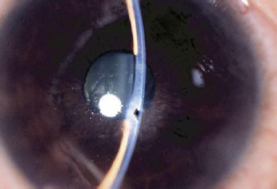
A 41-year-old man developed a small paracentral corneal perforation (0.5 mm in size) in the right eye, while the treating physician attempted to remove the residual rust ring after removal of a piece of metallic foreign body.
The eye was initially managed with a bandage soft contact lens to ameliorate the aqueous leakage; however, without success. Iatrogenic iris incarceration of the wound was first induced, followed by application of cyanoacrylate tissue adhesive to the perforated site. As a result, the anterior chamber was immediately reformed and maintained. Complete corneal epithelialization of the perforation was achieved in 2 months without visual compromises.
Cyanoacrylate tissue adhesive with iatrogenic incarceration of the autologous iris was effective in treating this type of small corneal perforation. This technique is simple and potentially useful for small paracentral corneal perforations outside the visual axis and without good apposition.
当眼前房在短时间内无法恢复时,需对角膜穿孔进行手术干预。在此,我们报告一例使用自体虹膜嵌顿和组织粘合剂成功治疗小的角膜旁中央穿孔的病例。
一名41岁男性右眼出现一个小的角膜旁中央穿孔(大小为0.5毫米),当时治疗医生在取出一块金属异物后试图去除残留的铁锈环。
最初用绷带软性接触镜处理该眼以改善房水渗漏,但未成功。首先诱导伤口发生医源性虹膜嵌顿,随后在穿孔部位应用氰基丙烯酸酯组织粘合剂。结果,眼前房立即恢复并得以维持。穿孔处角膜上皮在2个月内完全愈合,且视力未受影响。
氰基丙烯酸酯组织粘合剂联合自体虹膜医源性嵌顿对治疗此类小的角膜穿孔有效。该技术简单,对于视轴外且对合不佳的小角膜旁中央穿孔可能有用。