Department of Nuclear Medicine, Ludwig-Maximilians-University, Klinikum Grosshadern, Marchioninistr 15, Munich, 81377, Germany.
EJNMMI Res. 2012 Aug 3;2(1):43. doi: 10.1186/2191-219X-2-43.
We performed an initial evaluation of non-invasive ECG-gated [18 F]FDG-positron emission tomography (FDG-PET) for serial measurements of left ventricular volumes and function in murine models of dilated (DCM) and ischemic cardiomyopathy (ICM), and then tested the effect of erythropoietin (EPO) treatment on DCM mice in a preliminary FDG-PET therapy monitoring study.
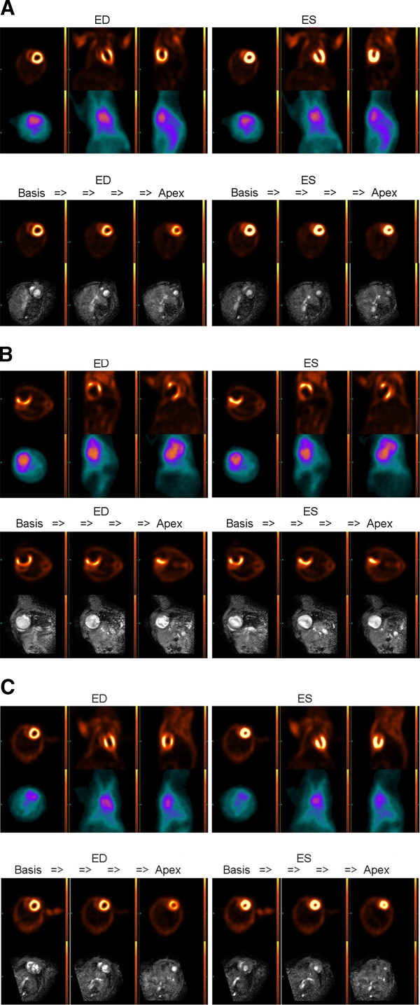
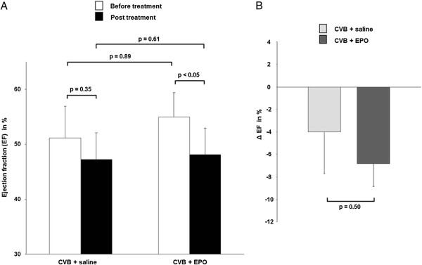
Mice developed DCM 8 weeks after injection with Coxsackievirus B3 (CVB3), whereas ICM was induced by ligation of the left anterior descending artery. LV volumes (EDV and ESV) and the ejection fraction (LVEF) of DCM, ICM and healthy control mice were measured by FDG-PET and compared with reference standard results obtained with 1.5 T magnetic resonance imaging (MRI). In the subsequent monitoring study, LVEF of DCM mice was evaluated by FDG-PET at baseline, and after 4 weeks of treatment, with EPO or saline.
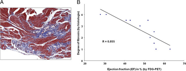
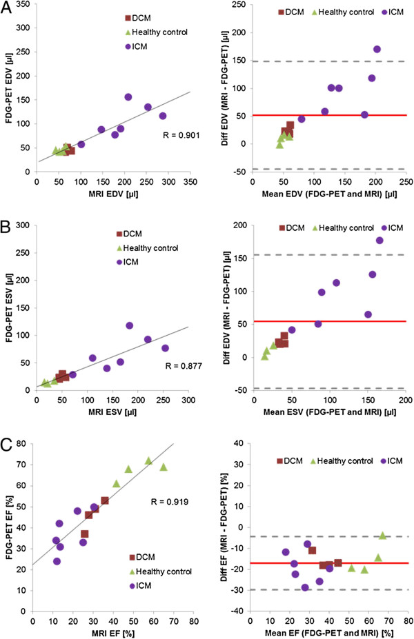
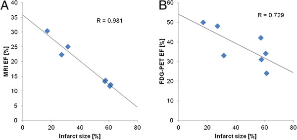
LV volumes and the LVEF as measured by FDG-PET correlated significantly with the MRI results. These correlations were higher in healthy and DCM mice than in ICM mice, in which LVEF measurements were somewhat compromised by absence of FDG uptake in the area of infarction. LV volumes (EDV and ESV) were systematically underestimated by FDG-PET, with net bias such that LVEF measurements in both models of heart disease exceeded by 15% to 20% results obtained by MRI. In our subsequent monitoring study of DCM mice, we found a significant decrease of LVEF in the EPO group, but not in the saline-treated mice. Moreover, LVEF in the EPO and saline mice significantly correlated with histological scores of fibrosis.
LVEF estimated by ECG-gated FDG-PET significantly correlated with the reference standard MRI, most notably in healthy mice and mice with DCM. FDG-PET served for longitudinal monitoring of effects of EPO treatment in DCM mice.
我们对非侵入性心电图门控 [18F]FDG-正电子发射断层扫描(FDG-PET)进行了初步评估,以连续测量扩张型心肌病(DCM)和缺血性心肌病(ICM)的小鼠模型中的左心室容积和功能,然后在初步的 FDG-PET 治疗监测研究中测试了促红细胞生成素(EPO)治疗对 DCM 小鼠的影响。
注射柯萨奇病毒 B3(CVB3) 8 周后,小鼠发展为 DCM,而左前降支结扎则诱导 ICM。通过 FDG-PET 测量 DCM、ICM 和健康对照组小鼠的左心室容积(EDV 和 ESV)和射血分数(LVEF),并与 1.5 T 磁共振成像(MRI)获得的参考标准结果进行比较。在随后的监测研究中,DCM 小鼠的 LVEF 通过 FDG-PET 在基线和 4 周治疗后进行评估,治疗方法为 EPO 或盐水。
FDG-PET 测量的左心室容积和 LVEF 与 MRI 结果显著相关。在健康和 DCM 小鼠中,这些相关性高于 ICM 小鼠,在 ICM 小鼠中,由于梗塞区域内缺乏 FDG 摄取,LVEF 测量受到一定影响。FDG-PET 系统地低估了左心室容积(EDV 和 ESV),其净偏差使得两种心脏病模型的 LVEF 测量值比 MRI 结果高 15%至 20%。在我们随后对 DCM 小鼠的监测研究中,我们发现 EPO 组的 LVEF 显著下降,但生理盐水治疗组的 LVEF 没有下降。此外,EPO 和生理盐水治疗的 DCM 小鼠的 LVEF 与纤维化的组织学评分显著相关。
心电图门控 FDG-PET 估计的 LVEF 与参考标准 MRI 显著相关,在健康小鼠和 DCM 小鼠中最为明显。FDG-PET 用于 DCM 小鼠 EPO 治疗效果的纵向监测。