Faculty of Engineering, Institute of Nanotechnology and Advanced Materials, Bar Ilan University, Ramat Gan, Israel.
Int J Nanomedicine. 2012;7:4707-13. doi: 10.2147/IJN.S34157. Epub 2012 Aug 28.
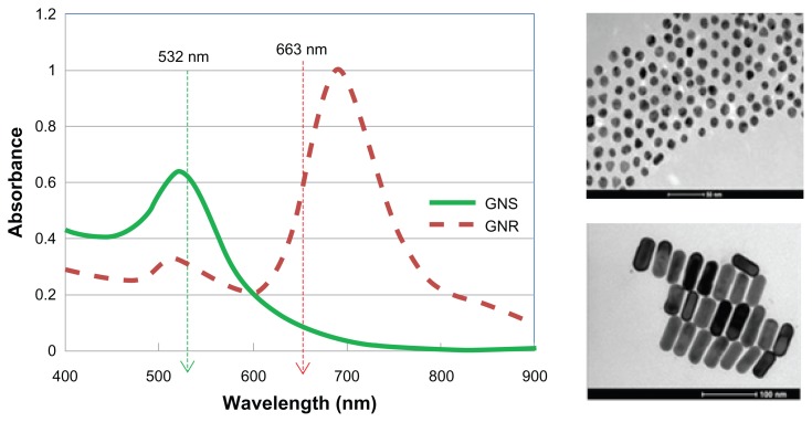
One of the critical problems in cancer management is local recurrence of disease. Between 20% and 30% of patients who undergo tumor resection surgery require reoperation due to incomplete excision. Currently, there are no validated methods for intraoperative tumor margin detection. In the present work, we demonstrate the potential use of gold nanoparticles (GNPs) as a novel contrast agent for photothermal molecular imaging of cancer.
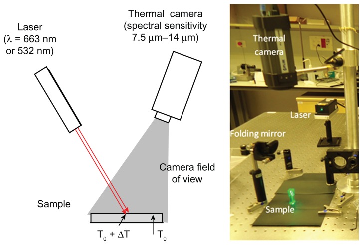
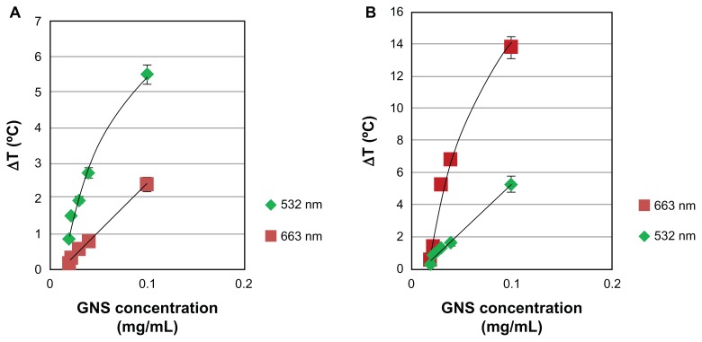
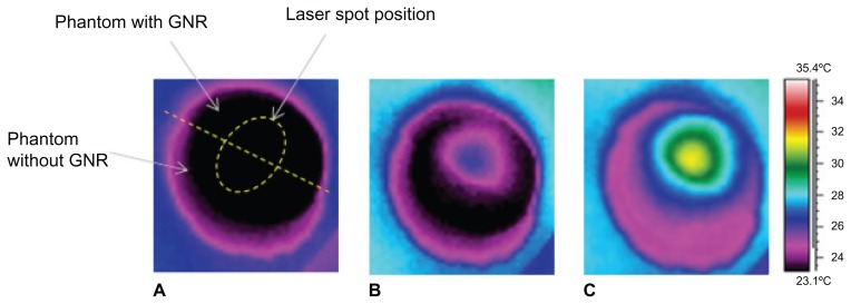
Phantoms containing different concentrations of GNPs were irradiated with continuous-wave laser and measured with a thermal imaging camera which detected the temperature field of the irradiated phantoms.
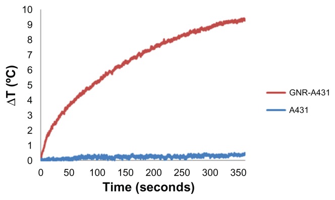
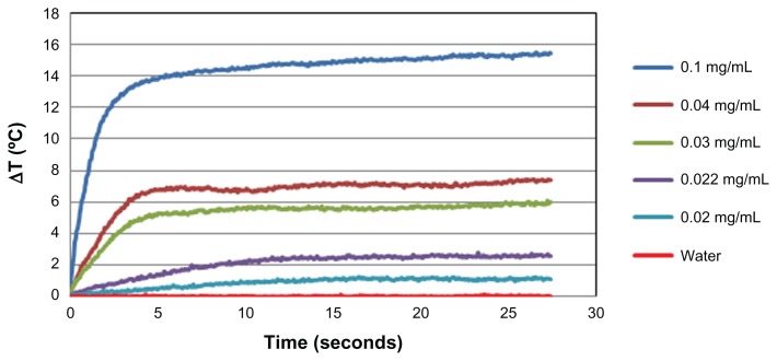
The results clearly demonstrate the ability to distinguish between cancerous cells specifically targeted with GNPs and normal cells. This technique, which allows highly sensitive discrimination between adjacent low GNP concentrations, will allow tumor margin detection while the temperature increases by only a few degrees Celsius (for GNPs in relevant biological concentrations).
We expect this real-time intraoperative imaging technique to assist surgeons in determining clear tumor margins and to maximize the extent of tumor resection while sparing normal background tissue.
癌症治疗中的一个关键问题是局部疾病复发。由于切除不完整, 20%至 30%的肿瘤切除术患者需要再次手术。目前,还没有用于术中肿瘤边界检测的验证方法。在本工作中,我们证明了金纳米粒子 (GNPs) 作为一种新型对比剂在癌症光热分子成像中的应用潜力。
含有不同浓度 GNPs 的体模用连续波激光照射,并使用热成像相机进行测量,该相机检测照射体模的温度场。
结果清楚地表明能够区分特异性靶向 GNPs 的癌细胞和正常细胞。这种技术可以在相邻低 GNPs 浓度之间进行高度敏感的区分,在温度仅升高几度的情况下(对于相关生物浓度的 GNPs)允许检测肿瘤边界。
我们期望这种实时术中成像技术能够帮助外科医生确定清晰的肿瘤边界,并最大限度地扩大肿瘤切除范围,同时保留正常背景组织。