Masedunskas Andrius, Milberg Oleg, Porat-Shliom Natalie, Sramkova Monika, Wigand Tim, Amornphimoltham Panomwat, Weigert Roberto
Intracellular Membrane Trafficking Unit; Oral and Pharyngeal Cancer Branch; National Institute of Dental and Craniofacial Research; National Institutes of Health; Bethesda, MD USA; Department of Biology; University of North Carolina at Chapel Hill; Chapel Hill, NC USA.
Intracellular Membrane Trafficking Unit; Oral and Pharyngeal Cancer Branch; National Institute of Dental and Craniofacial Research; National Institutes of Health; Bethesda, MD USA; Department of Chemical and Biochemical Engineering; Rutgers University; Piscataway, NJ USA; Department of Biomedical Engineering; Rutgers University; Piscataway, NJ USA.
Bioarchitecture. 2012 Sep-Oct;2(5):143-57. doi: 10.4161/bioa.21758. Epub 2012 Sep 1.
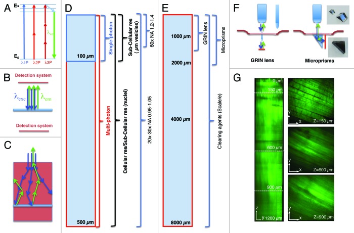
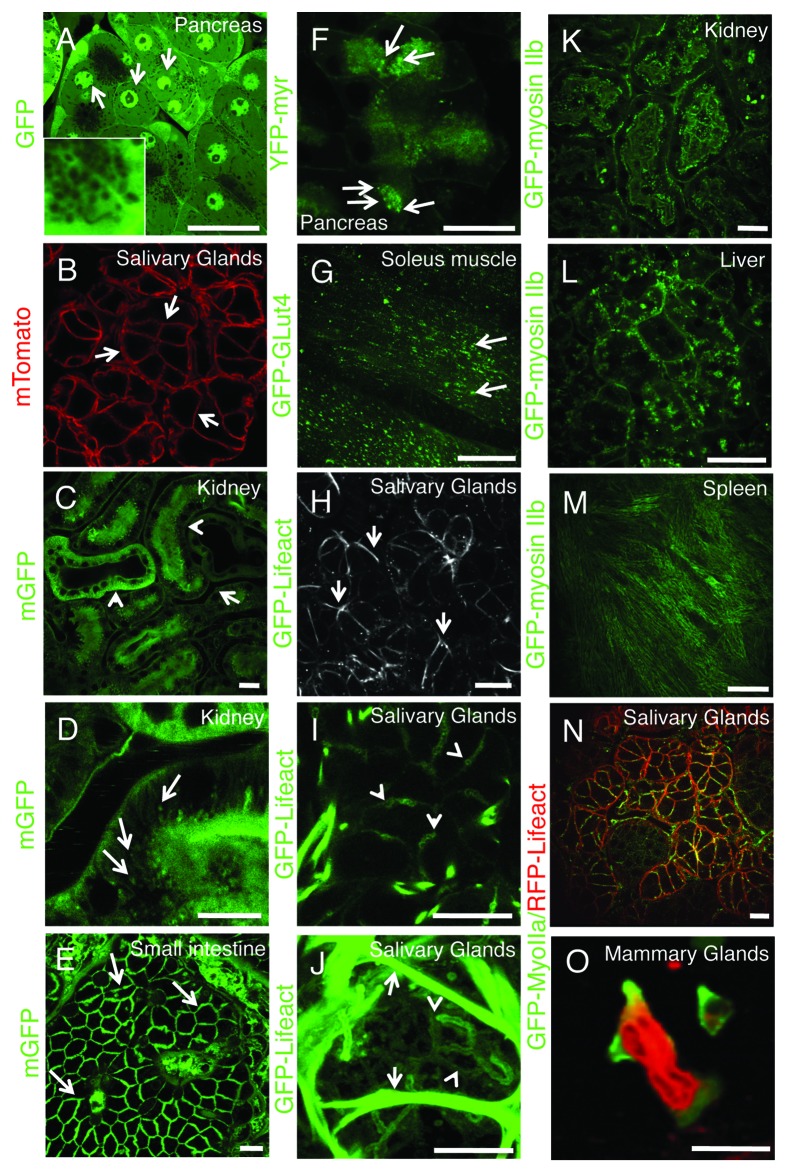
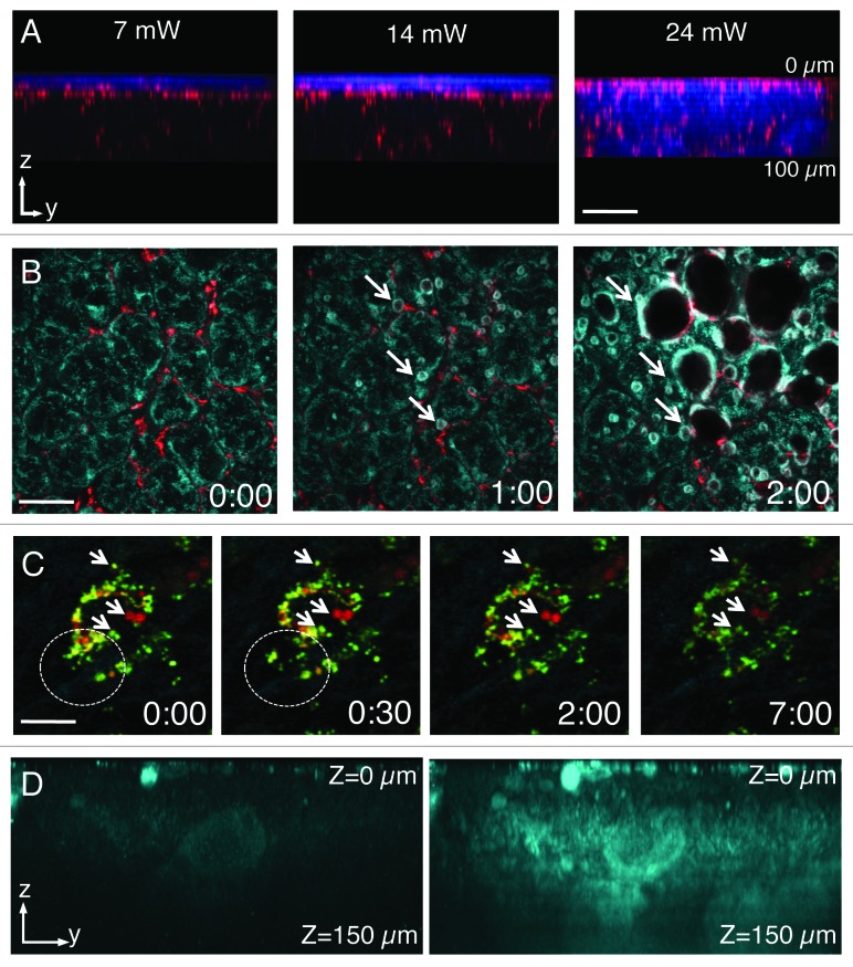
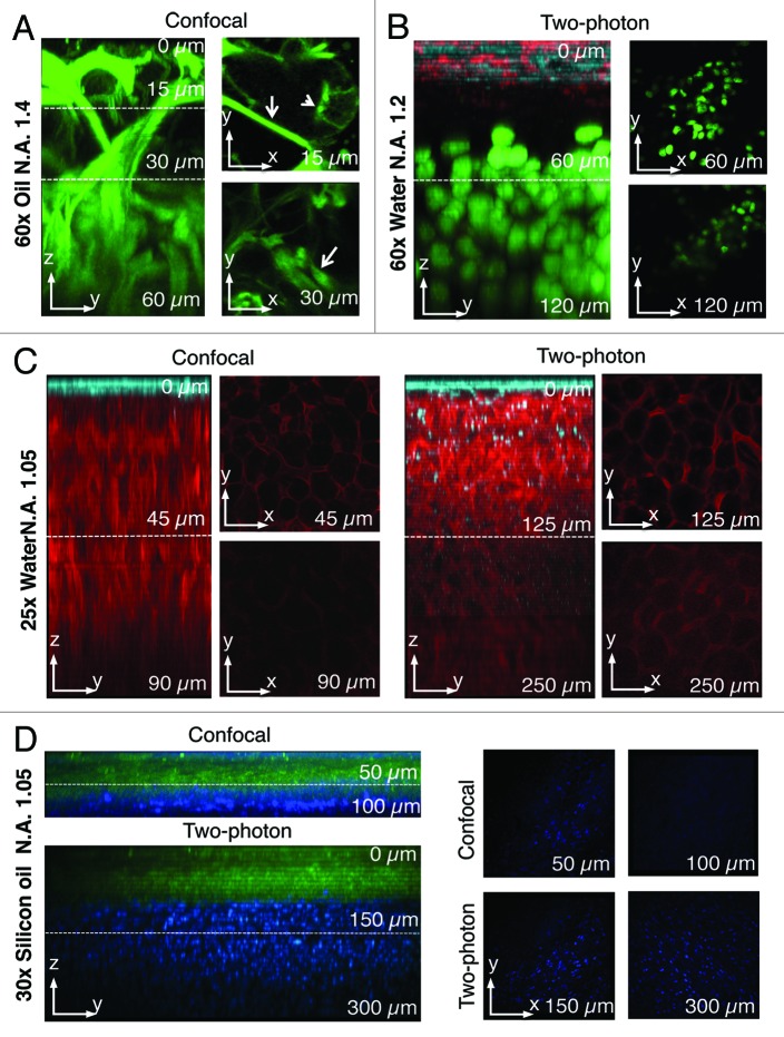
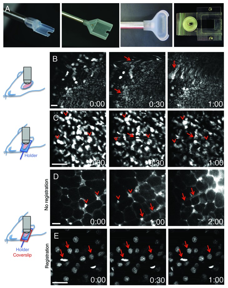
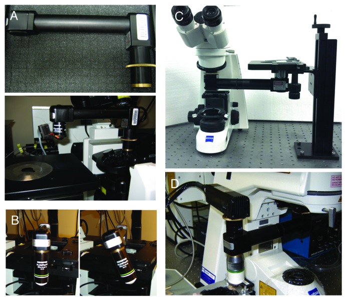
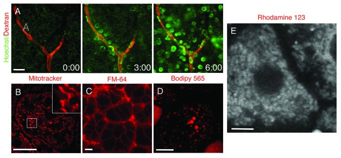
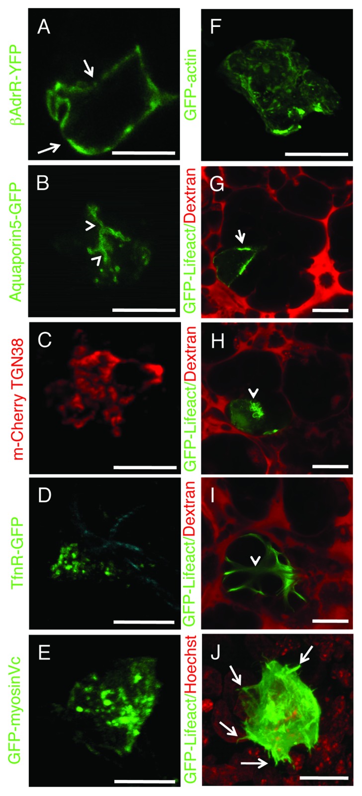
Intravital microscopy is an extremely powerful tool that enables imaging several biological processes in live animals. Recently, the ability to image subcellular structures in several organs combined with the development of sophisticated genetic tools has made possible extending this approach to investigate several aspects of cell biology. Here we provide a general overview of intravital microscopy with the goal of highlighting its potential and challenges. Specifically, this review is geared toward researchers that are new to intravital microscopy and focuses on practical aspects of carrying out imaging in live animals. Here we share the know-how that comes from first-hand experience, including topics such as choosing the right imaging platform and modality, surgery and stabilization techniques, anesthesia and temperature control. Moreover, we highlight some of the approaches that facilitate subcellular imaging in live animals by providing numerous examples of imaging selected organelles and the actin cytoskeleton in multiple organs.
活体显微镜检查是一种极其强大的工具,能够对活体动物体内的多种生物学过程进行成像。最近,在多个器官中对亚细胞结构进行成像的能力,再加上先进遗传工具的发展,使得将这种方法扩展到研究细胞生物学的多个方面成为可能。在此,我们对活体显微镜检查进行总体概述,旨在突出其潜力和挑战。具体而言,本综述面向初次接触活体显微镜检查的研究人员,重点关注在活体动物中进行成像的实际操作。我们在此分享来自第一手经验的实用知识,包括选择合适的成像平台和方式、手术及稳定技术、麻醉和温度控制等主题。此外,我们通过提供多个器官中选定细胞器和肌动蛋白细胞骨架成像的大量示例,突出了一些有助于在活体动物中进行亚细胞成像的方法。