Yamane Hideo, Sunami Kishiko, Iguchi Hiroyoshi, Sakamoto Hiramori, Imoto Toshio, Rask-Andersen Helge
Department of Otorhinolaryngology, Osaka City University Graduate School of Medicine, Japan.
Acta Otolaryngol. 2012 Oct;132(10):1054-60. doi: 10.3109/00016489.2012.680980.
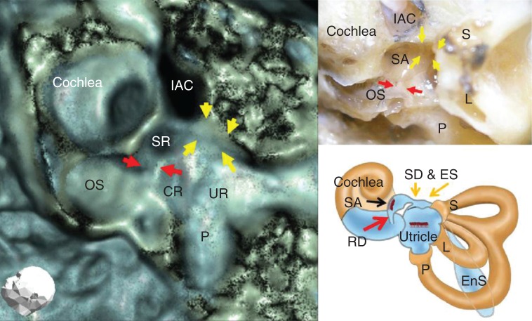
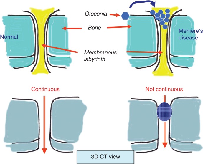
Significant reduced visualization of the reuniting duct (ductus reuniens; RD), saccular duct (SD) and endolymphatic sinus (ES) in Meniere's disease (MD) compared with normal control ears on three-dimensional (3D) CT imaging suggests the blockage of endolymphatic flow there with radiodense substances, which may be explained by dislodged otoconia from the saccule. These structures could be involved in the pathogenesis of MD.
This study was designed to visualize and assess the RD, SD and ES in patients with MD using 3D CT.
Sixty-two patients with a definite diagnose of unilateral MD, based on criteria proposed by the Committee on Hearing and Equilibrium of the American Academy of Otolaryngology-Head and Neck Surgery (AAO-HNS), were compared with contralateral ears and normal controls (26 ears) using 3D CT. The RD, SD and ES were scrutinized for patency on 3D CT images.
MD ears showed loss of continuity of the RD, SD and ES based on evaluation of 3D CT images, and differed significantly from normal healthy control ears (p < 0.01).
与正常对照耳相比,梅尼埃病(MD)患者三维(3D)CT成像显示联合管(再联合管;RD)、球囊导管(SD)和内淋巴窦(ES)的可视化显著降低,提示内淋巴流动在该处被放射性致密物质阻塞,这可能是由于球囊内耳石移位所致。这些结构可能参与了MD的发病机制。
本研究旨在使用3D CT对MD患者的RD、SD和ES进行可视化和评估。
根据美国耳鼻咽喉-头颈外科学会(AAO-HNS)听力与平衡委员会提出的标准,对62例确诊为单侧MD的患者进行3D CT检查,并与对侧耳和正常对照(26耳)进行比较。在3D CT图像上仔细检查RD、SD和ES的通畅情况。
根据3D CT图像评估,MD耳显示RD、SD和ES的连续性丧失,与正常健康对照耳有显著差异(p < 0.01)。