• 文献检索
  • 文档翻译
  • 深度研究
  • 学术资讯
  • Suppr Zotero 插件Zotero 插件
  • 邀请有礼
  • 套餐&价格
  • 历史记录
应用&插件
Suppr Zotero 插件Zotero 插件浏览器插件Mac 客户端Windows 客户端微信小程序
定价
高级版会员购买积分包购买API积分包
服务
文献检索文档翻译深度研究API 文档MCP 服务
关于我们
关于 Suppr公司介绍联系我们用户协议隐私条款
关注我们

Suppr 超能文献

核心技术专利:CN118964589B侵权必究
粤ICP备2023148730 号-1Suppr @ 2026

文献检索

告别复杂PubMed语法,用中文像聊天一样搜索,搜遍4000万医学文献。AI智能推荐,让科研检索更轻松。

立即免费搜索

文件翻译

保留排版,准确专业,支持PDF/Word/PPT等文件格式,支持 12+语言互译。

免费翻译文档

深度研究

AI帮你快速写综述,25分钟生成高质量综述,智能提取关键信息,辅助科研写作。

立即免费体验

定量评估冠心病患者的绝对心肌灌注:心血管磁共振与正电子发射断层扫描比较。

Quantification of absolute myocardial perfusion in patients with coronary artery disease: comparison between cardiovascular magnetic resonance and positron emission tomography.

机构信息

King's College London British Heart Foundation Centre of Excellence, National Institute of Health Research Biomedical Research Centre at Guy's and St. Thomas' NHS Foundation Trust, Wellcome Trust and Engineering and Physical Sciences Research Council Medical Engineering Centre, Division of Imaging Sciences, The Rayne Institute, St. Thomas' Hospital, London, United Kingdom.

出版信息

J Am Coll Cardiol. 2012 Oct 16;60(16):1546-55. doi: 10.1016/j.jacc.2012.05.052. Epub 2012 Sep 19.

DOI:10.1016/j.jacc.2012.05.052
PMID:22999722
原文链接:https://pmc.ncbi.nlm.nih.gov/articles/PMC7611225/
Abstract

OBJECTIVES

The aim of this study was to compare fully quantitative cardiovascular magnetic resonance (CMR) and positron emission tomography (PET) myocardial perfusion and myocardial perfusion reserve (MPR) measurements in patients with coronary artery disease (CAD).

BACKGROUND

Absolute quantification of myocardial perfusion and MPR with PET have proven diagnostic and prognostic roles in patients with CAD. Quantitative CMR perfusion imaging has been established more recently and has been validated against PET in normal hearts. However, there are no studies comparing fully quantitative CMR against PET perfusion imaging in patients with CAD.

METHODS

Forty-one patients with known or suspected CAD prospectively underwent quantitative (13)N-ammonia PET and CMR perfusion imaging before coronary angiography.

RESULTS

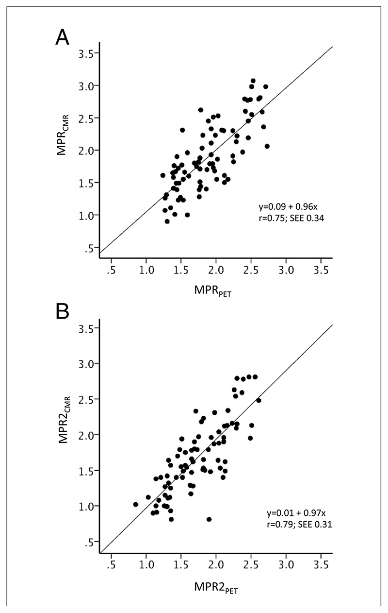
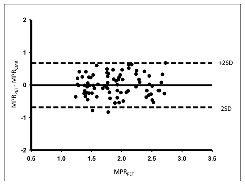
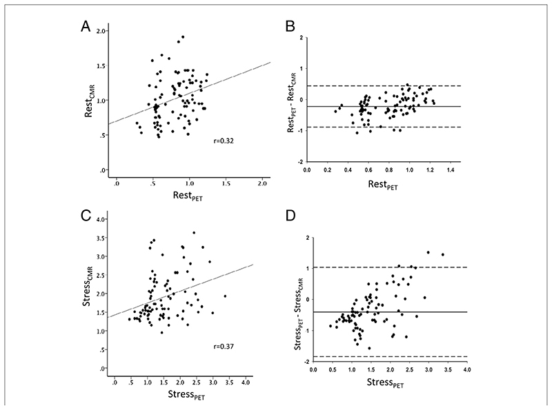
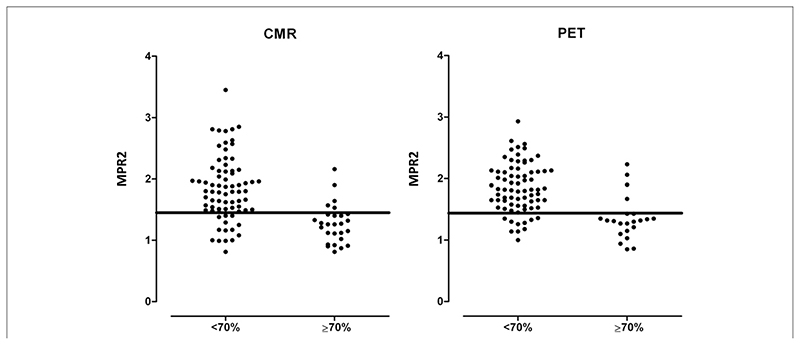
The CMR-derived MPR (MPR(CMR)) correlated well with PET-derived measurements (MPR(PET)) (r = 0.75, p < 0.0001). MPR(CMR) and MPR(PET) for the 2 lowest scoring segments in each coronary territory also correlated strongly (r = 0.79, p < 0.0001). Absolute CMR perfusion values correlated significantly, but weakly, with PET values both at rest (r = 0.32; p = 0.002) and during stress (r = 0.37; p < 0.0001). Area under the receiver-operating characteristic curve for MPR(PET) to detect significant CAD was 0.83 (95% confidence interval: 0.73 to 0.94) and for MPR(CMR) was 0.83 (95% confidence interval: 0.74 to 0.92). An MPR(PET) ≤1.44 predicted significant CAD with 82% sensitivity and 87% specificity, and MPR(CMR) ≤1.45 predicted significant CAD with 82% sensitivity and 81% specificity.

CONCLUSIONS

There is good correlation between MPR(CMR) and MPR(PET.) For the detection of significant CAD, MPR(PET) and MPR(CMR) seem comparable and very accurate. However, absolute perfusion values from PET and CMR are only weakly correlated; therefore, although quantitative CMR is clinically useful, further refinements are still required.

摘要

目的

本研究旨在比较冠状动脉疾病(CAD)患者的全定量心血管磁共振(CMR)和正电子发射断层扫描(PET)心肌灌注和心肌灌注储备(MPR)测量。

背景

PET 心肌灌注的绝对定量已被证明在 CAD 患者中有诊断和预后作用。定量 CMR 灌注成像较近才建立起来,并已在正常心脏中得到验证。然而,目前尚没有研究比较 CAD 患者的全定量 CMR 与 PET 灌注成像。

方法

41 例已知或疑似 CAD 的患者前瞻性地在冠状动脉造影前进行了定量(13)N-氨 PET 和 CMR 灌注成像。

结果

CMR 衍生的 MPR(MPR(CMR))与 PET 衍生的测量值(MPR(PET))密切相关(r = 0.75,p < 0.0001)。每个冠状动脉区域的 2 个评分最低节段的 MPR(CMR)和 MPR(PET)也具有很强的相关性(r = 0.79,p < 0.0001)。静息(r = 0.32;p = 0.002)和应激(r = 0.37;p < 0.0001)时,绝对 CMR 灌注值与 PET 值显著相关,但相关性较弱。MPR(PET)检测有意义的 CAD 的受试者工作特征曲线下面积为 0.83(95%置信区间:0.73 至 0.94),MPR(CMR)为 0.83(95%置信区间:0.74 至 0.92)。MPR(PET)≤1.44 预测有意义的 CAD 的敏感性为 82%,特异性为 87%,而 MPR(CMR)≤1.45 预测有意义的 CAD 的敏感性为 82%,特异性为 81%。

结论

MPR(CMR)与 MPR(PET)之间存在良好的相关性。对于检测有意义的 CAD,MPR(PET)和 MPR(CMR)似乎具有可比性,且非常准确。然而,PET 和 CMR 的绝对灌注值相关性较弱;因此,尽管定量 CMR 在临床上有用,但仍需要进一步改进。

https://cdn.ncbi.nlm.nih.gov/pmc/blobs/7824/7611225/f7d6f01a91a0/EMS129269-f005.jpg
https://cdn.ncbi.nlm.nih.gov/pmc/blobs/7824/7611225/b84598b49941/EMS129269-f001.jpg
https://cdn.ncbi.nlm.nih.gov/pmc/blobs/7824/7611225/b056bf77180d/EMS129269-f002.jpg
https://cdn.ncbi.nlm.nih.gov/pmc/blobs/7824/7611225/3d6de3f0935f/EMS129269-f003.jpg
https://cdn.ncbi.nlm.nih.gov/pmc/blobs/7824/7611225/068cb3d5667b/EMS129269-f004.jpg
https://cdn.ncbi.nlm.nih.gov/pmc/blobs/7824/7611225/f7d6f01a91a0/EMS129269-f005.jpg

相似文献

1
Quantification of absolute myocardial perfusion in patients with coronary artery disease: comparison between cardiovascular magnetic resonance and positron emission tomography.定量评估冠心病患者的绝对心肌灌注:心血管磁共振与正电子发射断层扫描比较。
J Am Coll Cardiol. 2012 Oct 16;60(16):1546-55. doi: 10.1016/j.jacc.2012.05.052. Epub 2012 Sep 19.
2
Fully automated pixel-wise quantitative CMR-myocardial perfusion with CMR-coronary angiography to detect hemodynamically significant coronary artery disease.应用 CMR 冠状动脉造影全自动像素级定量 CMR 心肌灌注成像检测有血流动力学意义的冠状动脉疾病。
Eur Radiol. 2023 Oct;33(10):7238-7249. doi: 10.1007/s00330-023-09689-8. Epub 2023 May 5.
3
Fully quantitative cardiovascular magnetic resonance myocardial perfusion ready for clinical use: a comparison between cardiovascular magnetic resonance imaging and positron emission tomography.全面定量心血管磁共振心肌灌注成像准备临床应用:心血管磁共振成像与正电子发射断层扫描比较。
J Cardiovasc Magn Reson. 2017 Oct 19;19(1):78. doi: 10.1186/s12968-017-0388-9.
4
Influence of the arterial input sampling location on the diagnostic accuracy of cardiovascular magnetic resonance stress myocardial perfusion quantification.动脉输入采样位置对心血管磁共振应激心肌灌注定量诊断准确性的影响。
J Cardiovasc Magn Reson. 2021 Mar 29;23(1):35. doi: 10.1186/s12968-021-00733-4.
5
Diagnostic Performance of Fully Automated Pixel-Wise Quantitative Myocardial Perfusion Imaging by Cardiovascular Magnetic Resonance.心血管磁共振全自动像素定量心肌灌注成像的诊断性能。
JACC Cardiovasc Imaging. 2018 May;11(5):697-707. doi: 10.1016/j.jcmg.2018.01.005. Epub 2018 Feb 14.
6
Diagnostic performance of quantitative perfusion cardiac magnetic resonance imaging in patients with prior coronary artery disease.定量灌注心脏磁共振成像在既往有冠状动脉疾病患者中的诊断性能
Eur Heart J Cardiovasc Imaging. 2025 Jan 31;26(2):207-217. doi: 10.1093/ehjci/jeae262.
7
Quantitative three-dimensional cardiovascular magnetic resonance myocardial perfusion imaging in systole and diastole.定量三维心血管磁共振心动周期收缩期和舒张期心肌灌注成像。
J Cardiovasc Magn Reson. 2014 Feb 24;16(1):19. doi: 10.1186/1532-429X-16-19.
8
Quantification of myocardial perfusion using cardiac magnetic resonance imaging correlates significantly to rubidium-82 positron emission tomography in patients with severe coronary artery disease: a preliminary study.利用心脏磁共振成像对心肌灌注进行定量分析,与严重冠状动脉疾病患者的铷-82正电子发射断层扫描结果显著相关:一项初步研究。
Eur J Radiol. 2014 Jul;83(7):1120-1128. doi: 10.1016/j.ejrad.2014.04.004. Epub 2014 Apr 16.
9
Qualitative assessments of myocardial ischemia by cardiac MRI and coronary stenosis by invasive coronary angiography in relation to quantitative perfusion by positron emission tomography in patients with known or suspected stable coronary artery disease.已知或疑似稳定型冠状动脉疾病患者的心脏 MRI 定性评估心肌缺血和有创性冠状动脉造影评估冠状动脉狭窄与正电子发射断层扫描定量评估灌注的关系。
J Nucl Cardiol. 2020 Dec;27(6):2351-2359. doi: 10.1007/s12350-018-01555-1. Epub 2018 Dec 10.
10
Automated Pixel-Wise Quantitative Myocardial Perfusion Mapping by CMR to Detect Obstructive Coronary Artery Disease and Coronary Microvascular Dysfunction: Validation Against Invasive Coronary Physiology.CMR 自动像素定量心肌灌注成像检测冠状动脉疾病和冠状动脉微血管功能障碍:与有创性冠状动脉生理学的对照验证。
JACC Cardiovasc Imaging. 2019 Oct;12(10):1958-1969. doi: 10.1016/j.jcmg.2018.12.022. Epub 2019 Feb 13.

引用本文的文献

1
Advanced Cardiac Magnetic Resonance Imaging for Assessment of Obstructive Coronary Artery Disease - ADVOCATE-CMR Study Rationale and Design.用于评估阻塞性冠状动脉疾病的高级心脏磁共振成像——ADVOCATE-CMR研究原理与设计
J Cardiovasc Magn Reson. 2025 Apr 25:101900. doi: 10.1016/j.jocmr.2025.101900.
2
Prevalence and prognostic significance of reduced myocardial perfusion reserve in diabetic heart failure with preserved ejection fraction using quantitative perfusion cardiac magnetic resonance.使用定量灌注心脏磁共振成像评估射血分数保留的糖尿病心力衰竭患者心肌灌注储备降低的患病率及预后意义
Eur Radiol. 2025 Mar 5. doi: 10.1007/s00330-025-11474-8.
3

本文引用的文献

1
Imaging in the management of ischemic cardiomyopathy: special focus on magnetic resonance.缺血性心肌病的影像学管理:特别关注磁共振。
J Am Coll Cardiol. 2012 Jan 24;59(4):359-70. doi: 10.1016/j.jacc.2011.08.076.
2
Clinical value of absolute quantification of myocardial perfusion with (15)O-water in coronary artery disease.(15)O-水心肌灌注绝对定量在冠心病中的临床价值。
Circ Cardiovasc Imaging. 2011 Nov;4(6):678-84. doi: 10.1161/CIRCIMAGING.110.960732. Epub 2011 Sep 16.
3
Development of a universal dual-bolus injection scheme for the quantitative assessment of myocardial perfusion cardiovascular magnetic resonance.
Quantitative perfusion by cardiac magnetic resonance imaging reveals compromised myocardial perfusion in patients with angina with non-obstructive coronary artery disease.
心脏磁共振成像定量灌注显示,非阻塞性冠状动脉疾病所致心绞痛患者存在心肌灌注受损。
Clin Res Cardiol. 2025 Feb 18. doi: 10.1007/s00392-025-02606-7.
4
Diagnostic performance of quantitative perfusion cardiac magnetic resonance imaging in patients with prior coronary artery disease.定量灌注心脏磁共振成像在既往有冠状动脉疾病患者中的诊断性能
Eur Heart J Cardiovasc Imaging. 2025 Jan 31;26(2):207-217. doi: 10.1093/ehjci/jeae262.
5
Coronary microvascular dysfunction in autoimmune rheumatic diseases: beyond coronary flow velocity reserve.自身免疫性风湿性疾病中的冠状动脉微血管功能障碍:超越冠状动脉血流储备
Front Cardiovasc Med. 2024 Aug 21;11:1372703. doi: 10.3389/fcvm.2024.1372703. eCollection 2024.
6
Cardiovascular Magnetic Resonance for the Evaluation of Arrhythmogenic Substrates in Patients with Systemic Autoimmunity: An Update.心血管磁共振成像在系统性自身免疫性疾病患者致心律失常基质评估中的应用:最新进展
Rev Cardiovasc Med. 2023 Oct 12;24(10):290. doi: 10.31083/j.rcm2410290. eCollection 2023 Oct.
7
Impact of Coronary Microvascular Dysfunction in Takotsubo Syndrome: Cause, Consequence or Both?冠状动脉微血管功能障碍在应激性心肌病中的作用:原因、后果还是两者皆有?
Rev Cardiovasc Med. 2024 May 11;25(5):163. doi: 10.31083/j.rcm2505163. eCollection 2024 May.
8
Clinical implementation of a fully automated quantitative perfusion cardiovascular magnetic resonance imaging workflow with a simplified dual-bolus contrast administration scheme.临床应用全自动定量灌注心血管磁共振成像工作流程,采用简化的双对比剂给药方案。
Sci Rep. 2024 Apr 26;14(1):9665. doi: 10.1038/s41598-024-60503-x.
9
High-resolution free-breathing automated quantitative myocardial perfusion by cardiovascular magnetic resonance for the detection of functionally significant coronary artery disease.心血管磁共振高分辨率自由呼吸自动定量心肌灌注成像检测有功能意义的冠状动脉疾病。
Eur Heart J Cardiovasc Imaging. 2024 Jun 28;25(7):914-925. doi: 10.1093/ehjci/jeae084.
10
Positron emission tomography and its role in the assessment of vulnerable plaques in comparison to other imaging modalities.正电子发射断层扫描及其与其他成像方式相比在易损斑块评估中的作用。
Front Med (Lausanne). 2024 Feb 15;10:1293848. doi: 10.3389/fmed.2023.1293848. eCollection 2023.
开发一种通用的双对比剂注射方案,用于定量评估心肌灌注心血管磁共振。
J Cardiovasc Magn Reson. 2011 May 24;13(1):28. doi: 10.1186/1532-429X-13-28.
4
High-resolution magnetic resonance myocardial perfusion imaging at 3.0-Tesla to detect hemodynamically significant coronary stenoses as determined by fractional flow reserve.3.0 特斯拉高分辨率磁共振心肌灌注成像检测血流储备分数测定的有意义的冠状动脉狭窄。
J Am Coll Cardiol. 2011 Jan 4;57(1):70-5. doi: 10.1016/j.jacc.2010.09.019.
5
Assessment of advanced coronary artery disease: advantages of quantitative cardiac magnetic resonance perfusion analysis.评估严重冠状动脉疾病:定量心脏磁共振灌注分析的优势。
J Am Coll Cardiol. 2010 Aug 10;56(7):561-9. doi: 10.1016/j.jacc.2010.02.061.
6
Relationship between regional myocardial oxygenation and perfusion in patients with coronary artery disease: insights from cardiovascular magnetic resonance and positron emission tomography.冠心病患者心肌氧合与灌注的关系:心血管磁共振和正电子发射断层扫描的见解。
Circ Cardiovasc Imaging. 2010 Jan;3(1):32-40. doi: 10.1161/CIRCIMAGING.109.860148. Epub 2009 Nov 17.
7
Long-term prognostic value of 13N-ammonia myocardial perfusion positron emission tomography added value of coronary flow reserve.13N-氨心肌灌注正电子发射断层扫描中冠状动脉血流储备增加值的长期预后价值。
J Am Coll Cardiol. 2009 Jul 7;54(2):150-6. doi: 10.1016/j.jacc.2009.02.069.
8
Comparison of positron emission tomography measurement of adenosine-stimulated absolute myocardial blood flow versus relative myocardial tracer content for physiological assessment of coronary artery stenosis severity and location.正电子发射断层扫描测量腺苷刺激后的绝对心肌血流量与相对心肌示踪剂含量用于评估冠状动脉狭窄严重程度和位置的生理比较
JACC Cardiovasc Imaging. 2009 Jun;2(6):751-8. doi: 10.1016/j.jcmg.2009.04.004.
9
Quantification of myocardial blood flow will reform the detection of CAD.心肌血流量的定量分析将革新冠心病的检测方法。
J Nucl Cardiol. 2009 Jul-Aug;16(4):497-506. doi: 10.1007/s12350-009-9101-1. Epub 2009 Jun 3.
10
Myocardial perfusion quantitation with 15O-labelled water PET: high reproducibility of the new cardiac analysis software (Carimas).用15O标记水PET进行心肌灌注定量分析:新型心脏分析软件(Carimas)具有高重现性。
Eur J Nucl Med Mol Imaging. 2009 Oct;36(10):1594-602. doi: 10.1007/s00259-009-1143-8. Epub 2009 Apr 30.