Department of Pathophysiology, School of Medicine, Jinan University, Guangzhou, Guangdong, China.
PLoS One. 2012;7(10):e47351. doi: 10.1371/journal.pone.0047351. Epub 2012 Oct 15.
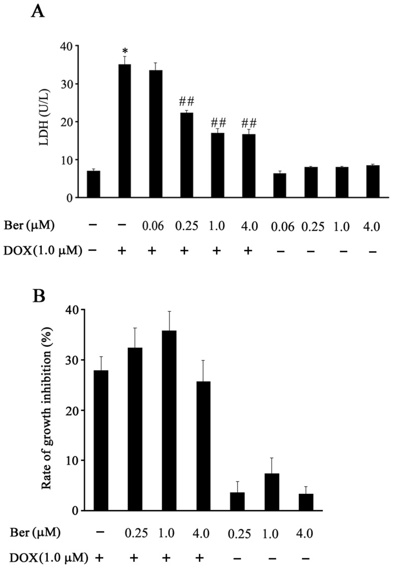
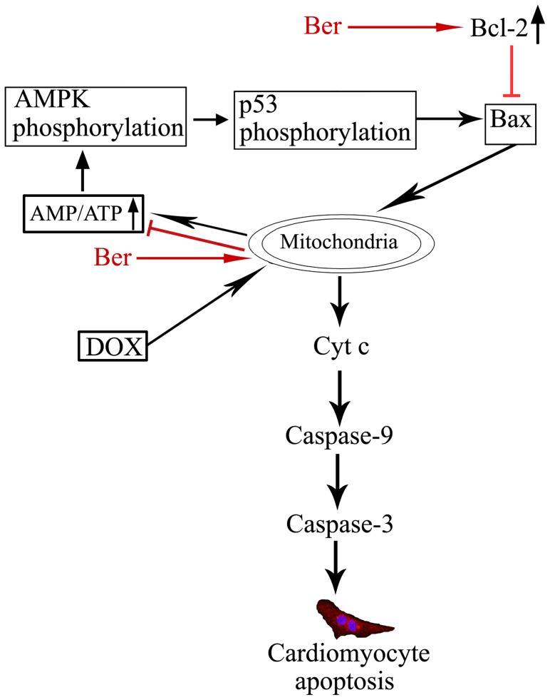
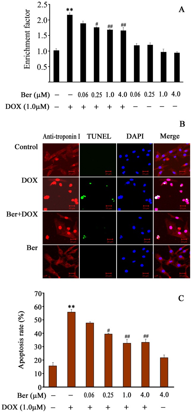
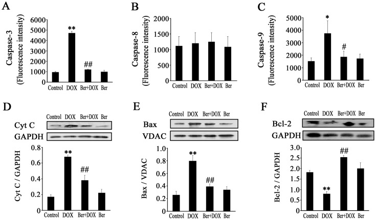
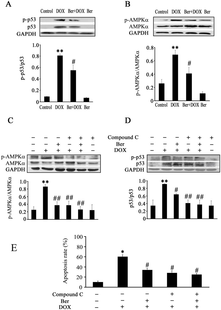
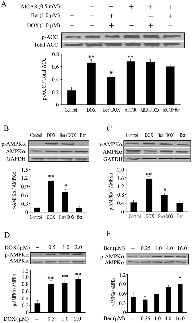
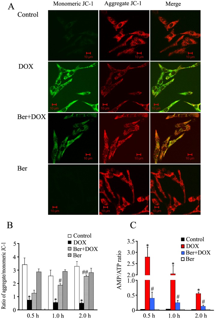
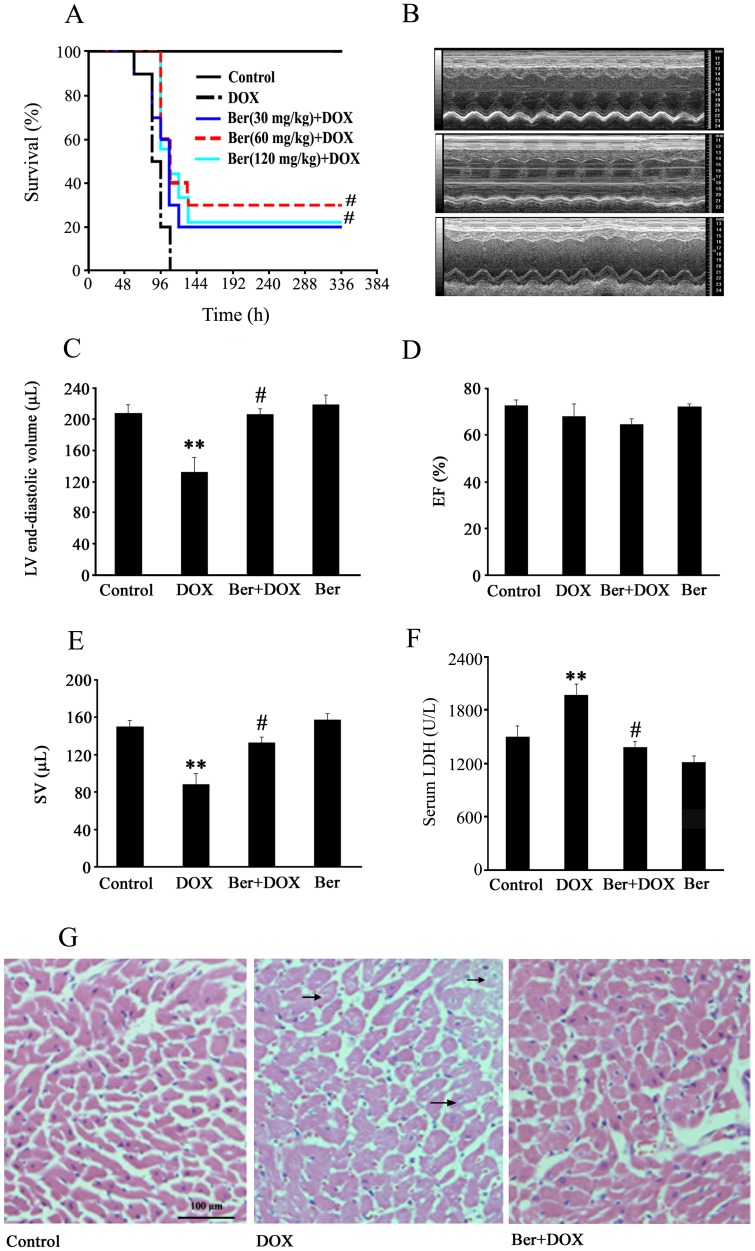
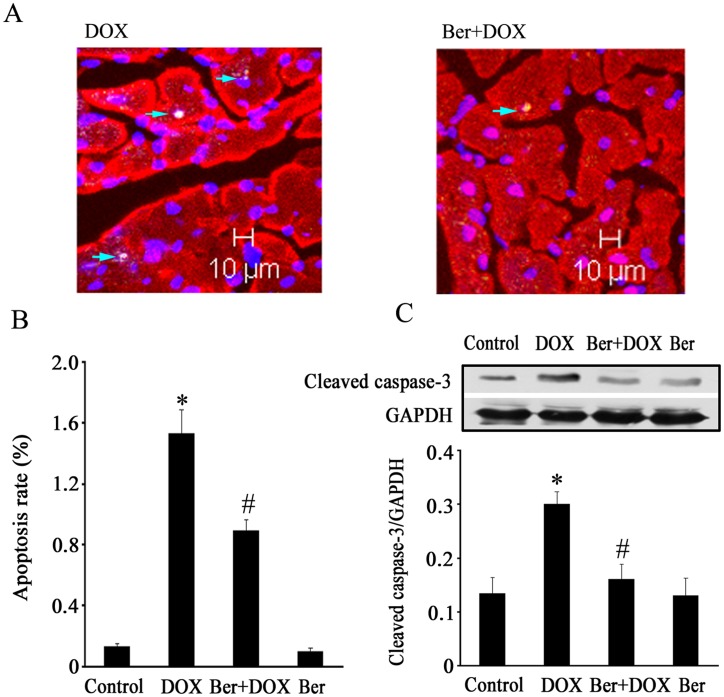
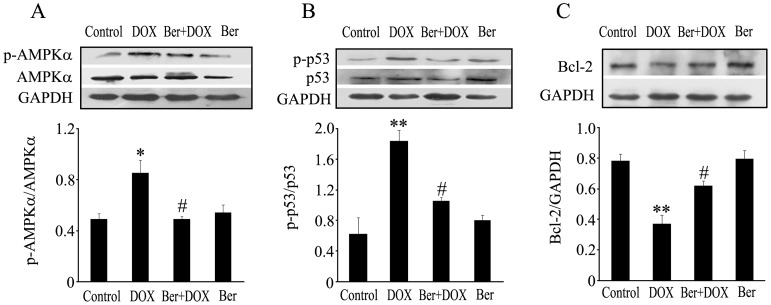
Cardiomyocyte apoptosis is an important event in doxorubicin (DOX)-induced cardiac injury. The aim of the present study was to investigate the protection of berberine (Ber) against DOX- triggered cardiomyocyte apoptosis in neonatal rat cardiomyocytes and rats. In neonatal rat cardiomyocytes, Ber attenuated DOX-induced cellular injury and apoptosis in a dose-dependent manner. However, Ber has no significant effect on viability of MCF-7 breast cancer cells treated with DOX. Ber reduced caspase-3 and caspase-9, but not caspase-8 activity in DOX-treated cardiomyocytes. Furthermore, Ber decreased adenosine monophosphate-activated protein kinase α (AMPKα) and p53 phosphorylation at 2 h, cytosolic cytochrome c and mitochondrial Bax levels and increased Bcl-2 level at 6 h in DOX-stimulated cardiomyocytes. Pretreatment with compound C, an AMPK inhibitor, also suppressed p53 phosphorylation and apoptosis in DOX-treated cardiomyocytes. DOX stimulation for 30 min led to a loss of mitochondrial membrane potential and a rise in the AMP/ATP ratio. Ber markedly reduced DOX-induced mitochondrial membrane potential loss and an increase in the AMP/ATP ratio at 1 h and 2 h post DOX exposure. In in vivo experiments, Ber significantly improved survival, increased stroke volume and attenuated myocardial injury in DOX-challenged rats. TUNEL and Western blot assays showed that Ber not only decreased myocardial apoptosis, caspase-3 activation, AMPKα and p53 phosphorylation, but also increased Bcl-2 expression in myocardium of rats exposed to DOX for 84 h. These findings indicate that Ber attenuates DOX-induced cardiomyocyte apoptosis via protecting mitochondria, inhibiting an increase in the AMP/ATP ratio and AMPKα phosphorylation as well as elevating Bcl-2 expression, which offer a novel mechanism responsible for protection of Ber against DOX-induced cardiomyopathy.
心肌细胞凋亡是多柔比星(DOX)诱导心脏损伤的一个重要事件。本研究旨在探讨小檗碱(Ber)对新生大鼠心肌细胞和大鼠 DOX 触发的心肌细胞凋亡的保护作用。在新生大鼠心肌细胞中,Ber 以剂量依赖的方式减弱 DOX 诱导的细胞损伤和凋亡。然而,Ber 对 DOX 处理的 MCF-7 乳腺癌细胞的活力没有显著影响。Ber 降低了 DOX 处理的心肌细胞中 caspase-3 和 caspase-9 的活性,但对 caspase-8 没有影响。此外,Ber 在 DOX 刺激的心肌细胞中,于 2 小时降低了腺苷单磷酸激活蛋白激酶α(AMPKα)和 p53 的磷酸化水平,于 6 小时降低了胞浆细胞色素 c 和线粒体 Bax 的水平,并增加了 Bcl-2 的水平。用 AMPK 抑制剂化合物 C 预处理也抑制了 DOX 处理的心肌细胞中的 p53 磷酸化和凋亡。DOX 刺激 30 分钟导致线粒体膜电位丧失和 AMP/ATP 比值升高。Ber 在 DOX 暴露后 1 小时和 2 小时显著减少了 DOX 诱导的线粒体膜电位丧失和 AMP/ATP 比值升高。在体内实验中,Ber 显著提高了 DOX 挑战大鼠的存活率,增加了每搏输出量,并减轻了心肌损伤。TUNEL 和 Western blot 检测表明,Ber 不仅降低了心肌细胞凋亡、caspase-3 活化、AMPKα 和 p53 的磷酸化,还增加了 DOX 暴露 84 小时的大鼠心肌中的 Bcl-2 表达。这些发现表明,Ber 通过保护线粒体、抑制 AMP/ATP 比值和 AMPKα 磷酸化的增加以及提高 Bcl-2 的表达来减轻 DOX 诱导的心肌细胞凋亡,为 Ber 对抗 DOX 诱导的心肌病提供了一种新的机制。