• 文献检索
  • 文档翻译
  • 深度研究
  • 学术资讯
  • Suppr Zotero 插件Zotero 插件
  • 邀请有礼
  • 套餐&价格
  • 历史记录
应用&插件
Suppr Zotero 插件Zotero 插件浏览器插件Mac 客户端Windows 客户端微信小程序
定价
高级版会员购买积分包购买API积分包
服务
文献检索文档翻译深度研究API 文档MCP 服务
关于我们
关于 Suppr公司介绍联系我们用户协议隐私条款
关注我们

Suppr 超能文献

核心技术专利:CN118964589B侵权必究
粤ICP备2023148730 号-1Suppr @ 2026

文献检索

告别复杂PubMed语法,用中文像聊天一样搜索,搜遍4000万医学文献。AI智能推荐,让科研检索更轻松。

立即免费搜索

文件翻译

保留排版,准确专业,支持PDF/Word/PPT等文件格式,支持 12+语言互译。

免费翻译文档

深度研究

AI帮你快速写综述,25分钟生成高质量综述,智能提取关键信息,辅助科研写作。

立即免费体验

CT 自动量化气胸

Automated quantification of pneumothorax in CT.

机构信息

Department of Radiology, Massachusetts General Hospital, 25 New Chardon Street, Suite 400B, Boston, MA 02114, USA.

出版信息

Comput Math Methods Med. 2012;2012:736320. doi: 10.1155/2012/736320. Epub 2012 Oct 3.

DOI:10.1155/2012/736320
PMID:23082091
原文链接:https://pmc.ncbi.nlm.nih.gov/articles/PMC3469107/
Abstract

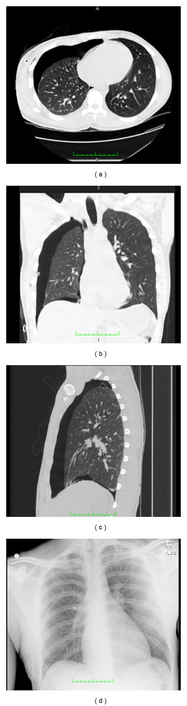
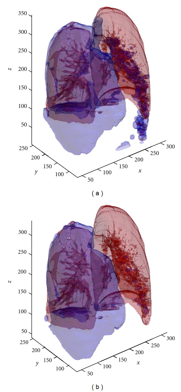
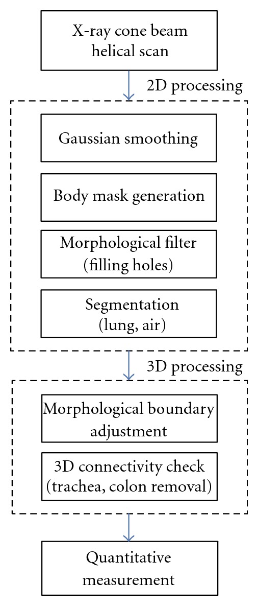
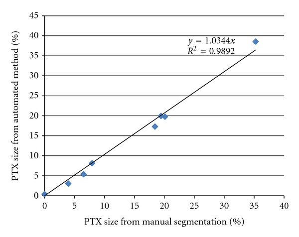
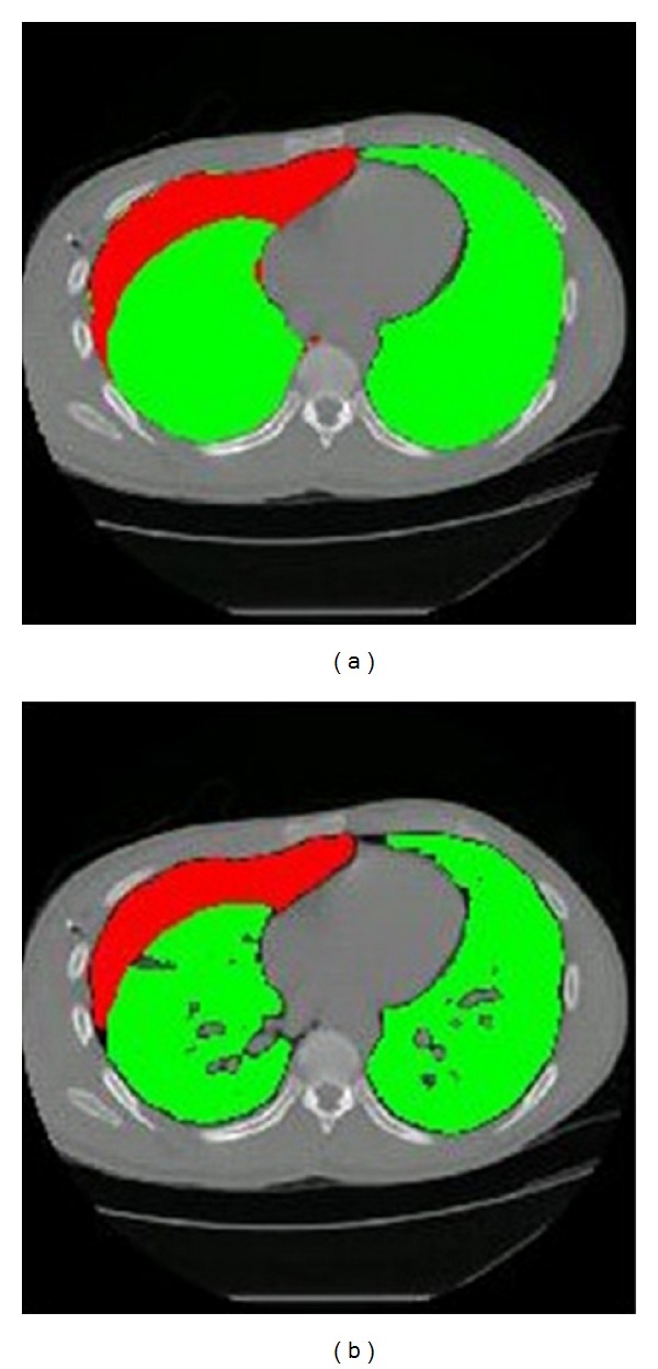
An automated, computer-aided diagnosis (CAD) algorithm for the quantification of pneumothoraces from Multidetector Computed Tomography (MDCT) images has been developed. Algorithm performance was evaluated through comparison to manual segmentation by expert radiologists. A combination of two-dimensional and three-dimensional processing techniques was incorporated to reduce required processing time by two-thirds (as compared to similar techniques). Volumetric measurements on relative pneumothorax size were obtained and the overall performance of the automated method shows an average error of just below 1%.

摘要

已经开发出一种用于从多排螺旋 CT(MDCT)图像定量气胸的自动化、计算机辅助诊断(CAD)算法。通过与专家放射科医生的手动分割进行比较来评估算法性能。结合了二维和三维处理技术,将所需的处理时间减少了三分之二(与类似技术相比)。获得了相对气胸大小的体积测量值,并且自动方法的整体性能显示平均误差仅略低于 1%。

https://cdn.ncbi.nlm.nih.gov/pmc/blobs/4e38/3469107/c63c7589a471/CMMM2012-736320.005.jpg
https://cdn.ncbi.nlm.nih.gov/pmc/blobs/4e38/3469107/20d92e3f6a5d/CMMM2012-736320.001.jpg
https://cdn.ncbi.nlm.nih.gov/pmc/blobs/4e38/3469107/6b5bc86a273a/CMMM2012-736320.002.jpg
https://cdn.ncbi.nlm.nih.gov/pmc/blobs/4e38/3469107/ed84dbfa7a5c/CMMM2012-736320.003.jpg
https://cdn.ncbi.nlm.nih.gov/pmc/blobs/4e38/3469107/f5063cb95027/CMMM2012-736320.004.jpg
https://cdn.ncbi.nlm.nih.gov/pmc/blobs/4e38/3469107/c63c7589a471/CMMM2012-736320.005.jpg

相似文献

1
Automated quantification of pneumothorax in CT.CT 自动量化气胸
Comput Math Methods Med. 2012;2012:736320. doi: 10.1155/2012/736320. Epub 2012 Oct 3.
2
MDCT for computerized volumetry of pneumothoraces in pediatric patients.MDCT 用于小儿气胸的计算机容积测量。
Acad Radiol. 2011 Mar;18(3):315-23. doi: 10.1016/j.acra.2010.11.008. Epub 2011 Jan 7.
3
Three-dimensional lung tumor segmentation from x-ray computed tomography using sparse field active models.基于稀疏域主动模型的 X 射线计算机断层扫描三维肺肿瘤分割。
Med Phys. 2012 Feb;39(2):851-65. doi: 10.1118/1.3676687.
4
MDCT quantification is the dominant parameter in decision-making regarding chest tube drainage for stable patients with traumatic pneumothorax.MDCT 定量是决定创伤性气胸稳定患者是否行胸腔引流管的主要参数。
Comput Med Imaging Graph. 2012 Jul;36(5):375-86. doi: 10.1016/j.compmedimag.2012.03.005. Epub 2012 May 4.
5
Automated segmentation and quantification of liver and spleen from CT images using normalized probabilistic atlases and enhancement estimation.利用归一化概率图谱和增强估计,对 CT 图像中的肝脏和脾脏进行自动分割和定量。
Med Phys. 2010 Feb;37(2):771-83. doi: 10.1118/1.3284530.
6
Geometrical model-based segmentation of the organs of sight on CT images.基于几何模型的CT图像上视觉器官分割
Med Phys. 2008 Feb;35(2):735-43. doi: 10.1118/1.2826557.
7
A minimum spanning forest based classification method for dedicated breast CT images.一种基于最小生成森林的专用乳腺CT图像分类方法。
Med Phys. 2015 Nov;42(11):6190-202. doi: 10.1118/1.4931958.
8
Relationship between coronary artery disease and epicardial adipose tissue quantification at cardiac CT: comparison between automatic volumetric measurement and manual bidimensional estimation.冠状动脉疾病与心脏 CT 下心外膜脂肪组织定量的关系:自动容积测量与手动二维估计的比较。
Acad Radiol. 2010 Jun;17(6):727-34. doi: 10.1016/j.acra.2010.01.015. Epub 2010 Apr 3.
9
Path planning for virtual bronchoscopy.虚拟支气管镜检查的路径规划
Conf Proc IEEE Eng Med Biol Soc. 2006;2006:156-9. doi: 10.1109/IEMBS.2006.259954.
10
A 3-D CAD tool for CT colonography.一种用于CT结肠成像的三维计算机辅助设计工具。
Annu Int Conf IEEE Eng Med Biol Soc. 2007;2007:3757-60. doi: 10.1109/IEMBS.2007.4353149.

引用本文的文献

1
Radiomics-based machine learning for automated detection of Pneumothorax in CT scans.基于影像组学的机器学习用于CT扫描中气胸的自动检测。
PLoS One. 2024 Dec 9;19(12):e0314988. doi: 10.1371/journal.pone.0314988. eCollection 2024.
2
Automatic and efficient pneumothorax segmentation from CT images using EFA-Net with feature alignment function.使用具有特征对齐功能的 EFA-Net 自动高效地从 CT 图像中分割气胸。
Sci Rep. 2023 Sep 15;13(1):15291. doi: 10.1038/s41598-023-42388-4.
3
Engineering and clinical use of artificial intelligence (AI) with machine learning and data science advancements: radiology leading the way for future.

本文引用的文献

1
Treatment of primary spontaneous pneumothorax.原发性自发性气胸的治疗
Curr Opin Pulm Med. 2009 Jul;15(4):376-9. doi: 10.1097/MCP.0b013e32832ae314.
2
MDCT for automated detection and measurement of pneumothoraces in trauma patients.多层螺旋CT用于创伤患者气胸的自动检测与测量。
AJR Am J Roentgenol. 2009 Mar;192(3):830-6. doi: 10.2214/AJR.08.1339.
3
Pneumothorax.气胸
随着机器学习和数据科学的进步,人工智能(AI)的工程应用与临床应用:放射学引领未来之路。
Ther Adv Urol. 2021 Sep 20;13:17562872211044880. doi: 10.1177/17562872211044880. eCollection 2021 Jan-Dec.
4
Deep learning detection and quantification of pneumothorax in heterogeneous routine chest computed tomography.深度学习在异质常规胸部 CT 中检测和量化气胸。
Eur Radiol Exp. 2020 Apr 17;4(1):26. doi: 10.1186/s41747-020-00152-7.
5
Anatomical locations of air for rapid diagnosis of pneumothorax in blunt trauma patients.在钝性创伤患者中快速诊断气胸的空气解剖位置。
World J Emerg Surg. 2019 Sep 2;14:44. doi: 10.1186/s13017-019-0263-0. eCollection 2019.
6
Current Applications and Future Impact of Machine Learning in Radiology.机器学习在放射学中的当前应用和未来影响。
Radiology. 2018 Aug;288(2):318-328. doi: 10.1148/radiol.2018171820. Epub 2018 Jun 26.
7
Computerized Diagnostic Assistant for the Automatic Detection of Pneumothorax on Ultrasound: A Pilot Study.用于超声自动检测气胸的计算机诊断助手:一项初步研究。
West J Emerg Med. 2016 Mar;17(2):209-15. doi: 10.5811/westjem.2016.1.28087. Epub 2016 Mar 2.
8
Evaluation of three pneumothorax size quantification methods on digitized chest X-ray films using medical-grade grayscale and consumer-grade color displays.使用医学级灰度显示器和消费级彩色显示器对数字化胸部X光片上的三种气胸大小量化方法进行评估。
J Digit Imaging. 2014 Apr;27(2):280-6. doi: 10.1007/s10278-013-9651-2.
Respiration. 2008;76(2):121-7. doi: 10.1159/000135932. Epub 2008 Jun 26.
4
Pneumothorax.气胸
Respirology. 2004 Jun;9(2):157-64. doi: 10.1111/j.1440-1843.2004.00577.x.
5
Spontaneous pneumothorax and its treatment.自发性气胸及其治疗
J Am Med Assoc. 1954 May 1;155(1):24-9. doi: 10.1001/jama.1954.03690190030009.
6
BTS guidelines for the management of spontaneous pneumothorax.英国胸科学会自发性气胸管理指南。
Thorax. 2003 May;58 Suppl 2(Suppl 2):ii39-52. doi: 10.1136/thorax.58.suppl_2.ii39.
7
Traumatic pneumothorax detection with thoracic US: correlation with chest radiography and CT--initial experience.超声检查对创伤性气胸的诊断:与胸部X线和CT的相关性——初步经验
Radiology. 2002 Oct;225(1):210-4. doi: 10.1148/radiol.2251011102.
8
A population-based study on pneumothorax in severely traumatized patients.一项针对严重创伤患者气胸的基于人群的研究。
J Trauma. 2001 Oct;51(4):677-82. doi: 10.1097/00005373-200110000-00009.
9
Management of spontaneous pneumothorax: an American College of Chest Physicians Delphi consensus statement.自发性气胸的管理:美国胸科医师学会德尔菲共识声明
Chest. 2001 Feb;119(2):590-602. doi: 10.1378/chest.119.2.590.
10
Evaluation of the utility of computed tomography in the initial assessment of the critical care patient with chest trauma.计算机断层扫描在胸部创伤重症监护患者初始评估中的效用评估。
Crit Care Med. 2000 May;28(5):1370-5. doi: 10.1097/00003246-200005000-00018.