Laboratory of Virology, Rocky Mountain Laboratories, NIAID, NIH, Hamilton, Montana, United States of America.
PLoS One. 2012;7(10):e47912. doi: 10.1371/journal.pone.0047912. Epub 2012 Oct 24.
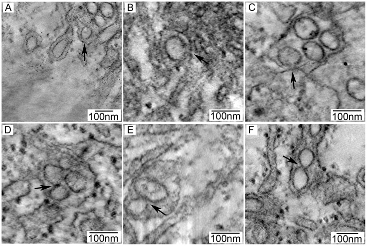
Tick-borne flaviviruses (TBFV) are sustained in nature through cycling between mammalian and tick hosts. In this study, we used African green monkey kidney cells (Vero) and Ixodes scapularis tick cells (ISE6) to compare virus-induced changes in mammalian and arthropod cells. Using confocal microscopy, transmission electron microscopy (TEM), and electron tomography (ET), we examined viral protein distribution and the ultrastructural changes that occur during TBFV infection. Within host cells, flaviviruses cause complex rearrangement of cellular membranes for the purpose of virus replication. Virus infection was accompanied by a marked expansion in endoplasmic reticulum (ER) staining and markers for TBFV replication were localized mainly to the ER in both cell lines. TEM of Vero cells showed membrane-bound vesicles enclosed in a network of dilated, anastomosing ER cisternae. Virions were seen within the ER and were sometimes in paracrystalline arrays. Tubular structures or elongated vesicles were occasionally noted. In acutely and persistently infected ISE6 cells, membrane proliferation and vesicles were also noted; however, the extent of membrane expansion and the abundance of vesicles were lower and no viral particles were observed. Tubular profiles were far more prevalent in persistently infected ISE6 cells than in acutely infected cells. By ET, tubular profiles, in persistently infected tick cells, had a cross-sectional diameter of 60-100 nm, reached up to 800 nm in length, were closed at the ends, and were often arranged in fascicle-like bundles, shrouded with ER membrane. Our experiments provide analysis of viral protein localization within the context of both mammalian and arthropod cell lines as well as both acute and persistent arthropod cell infection. Additionally, we show for the first time 3D flavivirus infection in a vector cell line and the first ET of persistent flavivirus infection.
蜱传黄病毒(TBFV)通过在哺乳动物和蜱宿主之间循环而在自然界中持续存在。在这项研究中,我们使用非洲绿猴肾细胞(Vero)和Ixodes scapularis 蜱细胞(ISE6)来比较病毒感染对哺乳动物和节肢动物细胞的影响。通过共聚焦显微镜、透射电子显微镜(TEM)和电子断层扫描(ET),我们检查了病毒蛋白的分布以及 TBFV 感染过程中发生的超微结构变化。在宿主细胞内,黄病毒会引起细胞膜的复杂重排,以进行病毒复制。病毒感染伴随着内质网(ER)染色的明显扩张,并且在两种细胞系中,TBFV 复制的标志物主要定位于 ER。Vero 细胞的 TEM 显示,膜结合的囊泡被扩张的、吻合的 ER 腔的网络所包围。病毒粒子在内质网内可见,有时呈准晶排列。偶尔还观察到管状结构或拉长的囊泡。在急性和持续性感染的 ISE6 细胞中,也观察到了膜增殖和囊泡;然而,膜扩张的程度和囊泡的丰度较低,并且没有观察到病毒颗粒。在持续性感染的 ISE6 细胞中,管状结构比急性感染细胞更为普遍。通过 ET,在持续性感染的蜱细胞中,管状结构的横截面直径为 60-100nm,长度可达 800nm,两端封闭,通常呈束状排列,被 ER 膜包裹。我们的实验提供了在哺乳动物和节肢动物细胞系以及急性和持续性节肢动物细胞感染的背景下对病毒蛋白定位的分析。此外,我们首次展示了在载体细胞系中 3D 黄病毒感染和首次 ET 持续性黄病毒感染。