Yoshikawa Hisao, Suzuki Makoto, Hashimoto Go, Kusunose Yukiko, Otsuka Takenori, Nakamura Masato, Sugi Kaoru
Division of Cardiovascular Medicine, Toho University Ohashi Medical Center, 2-17-6 Ohashi, Meguro-ku, Tokyo, Japan.
Cardiovasc Ultrasound. 2012 Nov 20;10:45. doi: 10.1186/1476-7120-10-45.
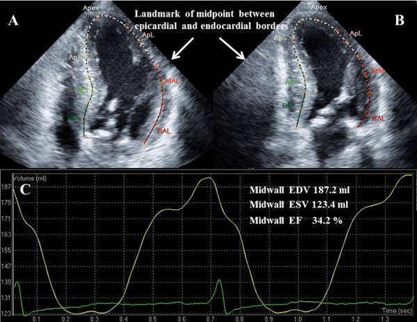
In patients with left ventricular hypertrophy (LVH), LV midwall fractional shortening (FS) is used as a measure of LV systolic performance that is more physiologically appropriate than conventional FS. For evaluation of LV volume and ejection fraction (EF), 2-dimensional (2D) echocardiography is more accurate than M-mode echocardiography. The purpose of this study was to assess systolic performance by midwall EF using 2D speckle tracking echocardiography (STE).
Sixty patients were enrolled in the study. Patients were divided into two groups with LVH (n = 30) and without LVH (control group, n = 30). LV systolic function was compared between the two groups and the relationships of left ventricular mass index (LVMI) with LV systolic parameters, including midwall EF, were investigated.
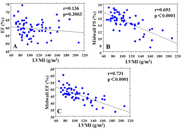
Midwall EF in the LVH group was significantly lower than that in the control group (42.8±4.4% vs. 48.1±4.1%, p <0.0001). Midwall FS was also significantly lower in the LVH group (13.4±2.8% vs. 16.1±1.5%, p <0.0001), but EF did not differ significantly between the two groups. There were significant correlations between midwall EF and LVMI (r=0.731, p <0.0001) and between midwall FS and LVMI (r=0.693, p <0.0001), with midwall EF having the higher correlation.
These results show that midwall EF can be determined using 2D STE. Midwall EF can be used to monitor LV systolic dysfunction, which is not possible with conventional EF. Evaluation of midwall EF may allow assessment of new parameters of LV systolic function in patients with LV geometric variability.
在左心室肥厚(LVH)患者中,左心室中层缩短分数(FS)被用作评估左心室收缩功能的指标,该指标在生理上比传统的FS更合适。对于左心室容积和射血分数(EF)的评估,二维(2D)超声心动图比M型超声心动图更准确。本研究的目的是使用2D斑点追踪超声心动图(STE)通过中层EF评估收缩功能。
60例患者纳入本研究。患者分为有LVH组(n = 30)和无LVH组(对照组,n = 30)。比较两组的左心室收缩功能,并研究左心室质量指数(LVMI)与包括中层EF在内的左心室收缩参数之间的关系。
LVH组的中层EF显著低于对照组(42.8±4.4% 对 48.1±4.1%,p <0.0001)。LVH组的中层FS也显著降低(13.4±2.8% 对 16.1±1.5%,p <0.0001),但两组之间的EF无显著差异。中层EF与LVMI之间存在显著相关性(r = 0.731,p <0.0001),中层FS与LVMI之间也存在显著相关性(r = 0.693,p <0.0001),中层EF的相关性更高。
这些结果表明,中层EF可以通过2D STE测定。中层EF可用于监测左心室收缩功能障碍,而传统EF无法做到这一点。对中层EF的评估可能有助于评估左心室几何形状变异患者左心室收缩功能的新参数。