• 文献检索
  • 文档翻译
  • 深度研究
  • 学术资讯
  • Suppr Zotero 插件Zotero 插件
  • 邀请有礼
  • 套餐&价格
  • 历史记录
应用&插件
Suppr Zotero 插件Zotero 插件浏览器插件Mac 客户端Windows 客户端微信小程序
定价
高级版会员购买积分包购买API积分包
服务
文献检索文档翻译深度研究API 文档MCP 服务
关于我们
关于 Suppr公司介绍联系我们用户协议隐私条款
关注我们

Suppr 超能文献

核心技术专利:CN118964589B侵权必究
粤ICP备2023148730 号-1Suppr @ 2026

文献检索

告别复杂PubMed语法,用中文像聊天一样搜索,搜遍4000万医学文献。AI智能推荐,让科研检索更轻松。

立即免费搜索

文件翻译

保留排版,准确专业,支持PDF/Word/PPT等文件格式,支持 12+语言互译。

免费翻译文档

深度研究

AI帮你快速写综述,25分钟生成高质量综述,智能提取关键信息,辅助科研写作。

立即免费体验

皮质多发性硬化病变的定量磁共振成像

Quantitative magnetic resonance imaging of cortical multiple sclerosis pathology.

作者信息

Tardif Christine L, Bedell Barry J, Eskildsen Simon F, Collins D Louis, Pike G Bruce

机构信息

McConnell Brain Imaging Centre, Montreal Neurological Institute, Montreal, Canada.

出版信息

Mult Scler Int. 2012;2012:742018. doi: 10.1155/2012/742018. Epub 2012 Nov 18.

DOI:10.1155/2012/742018
PMID:23213531
原文链接:https://pmc.ncbi.nlm.nih.gov/articles/PMC3506905/
Abstract

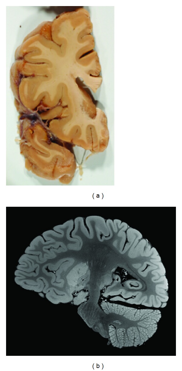
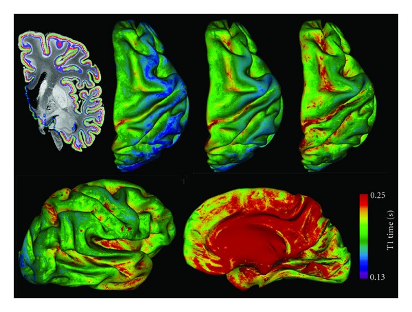
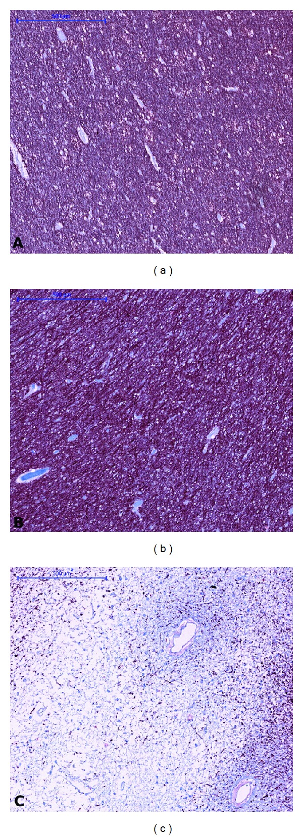
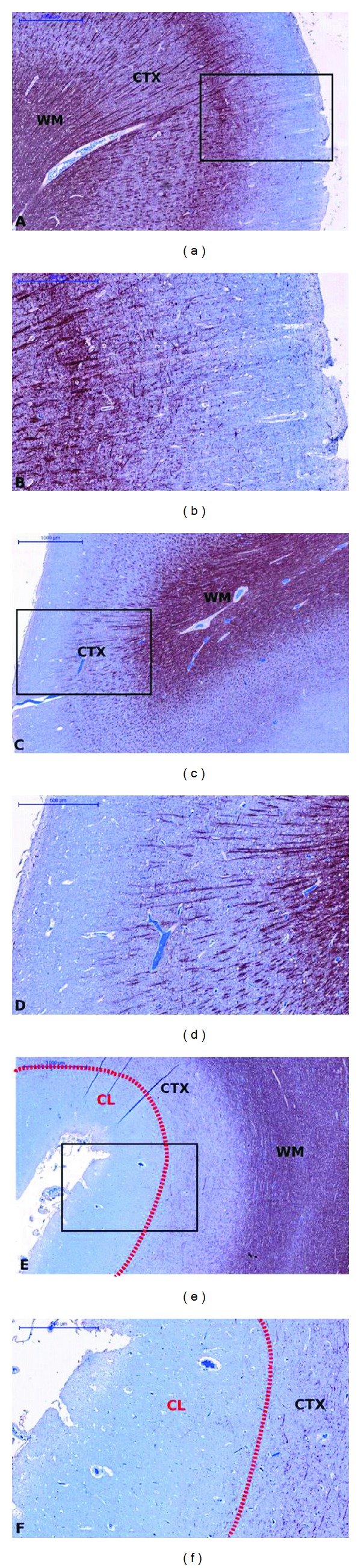
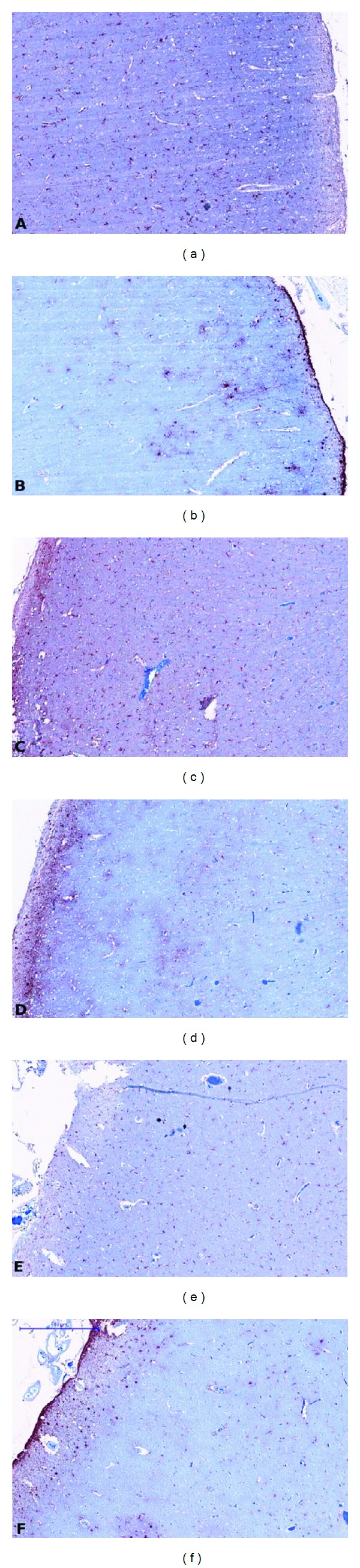
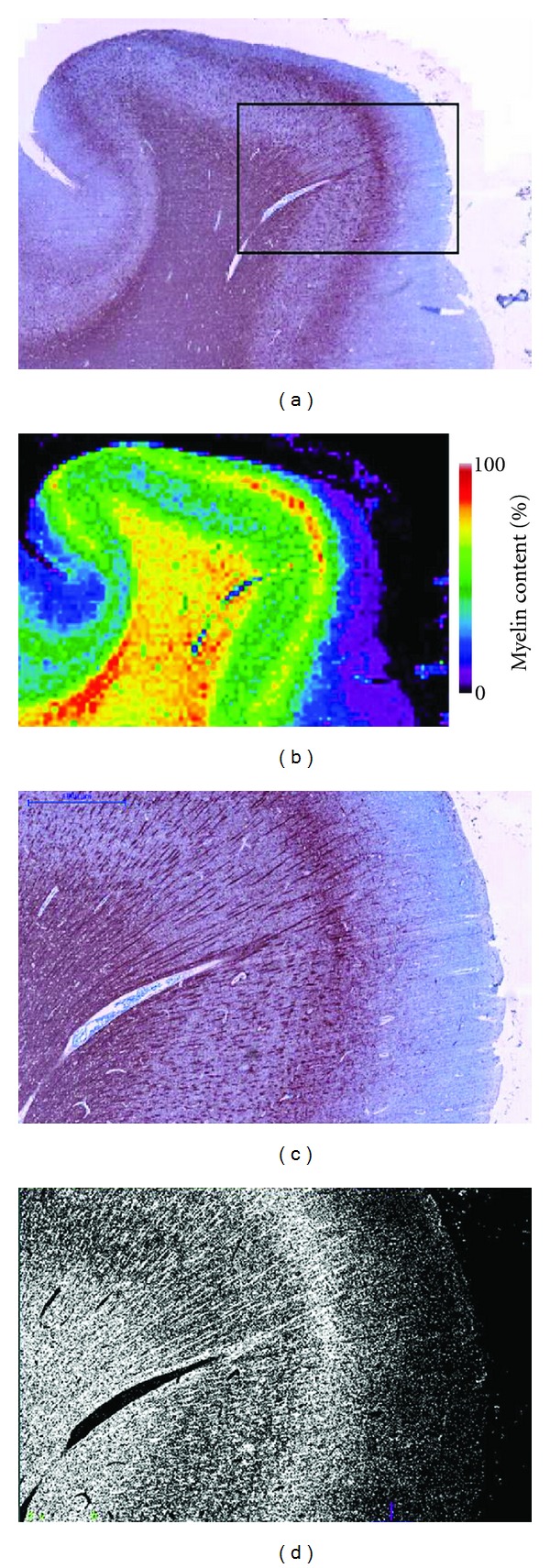
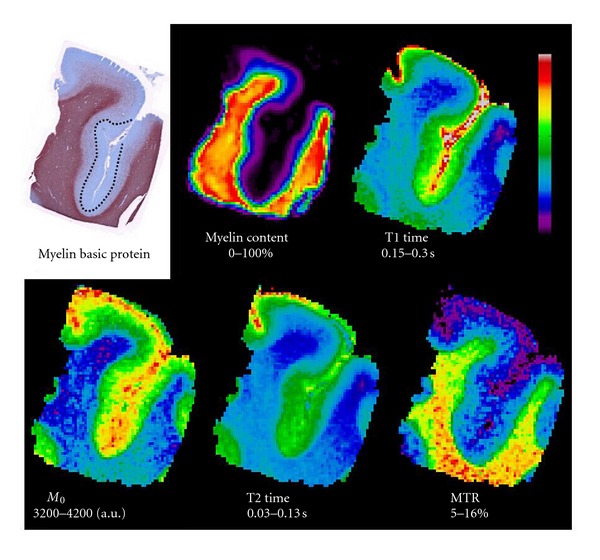
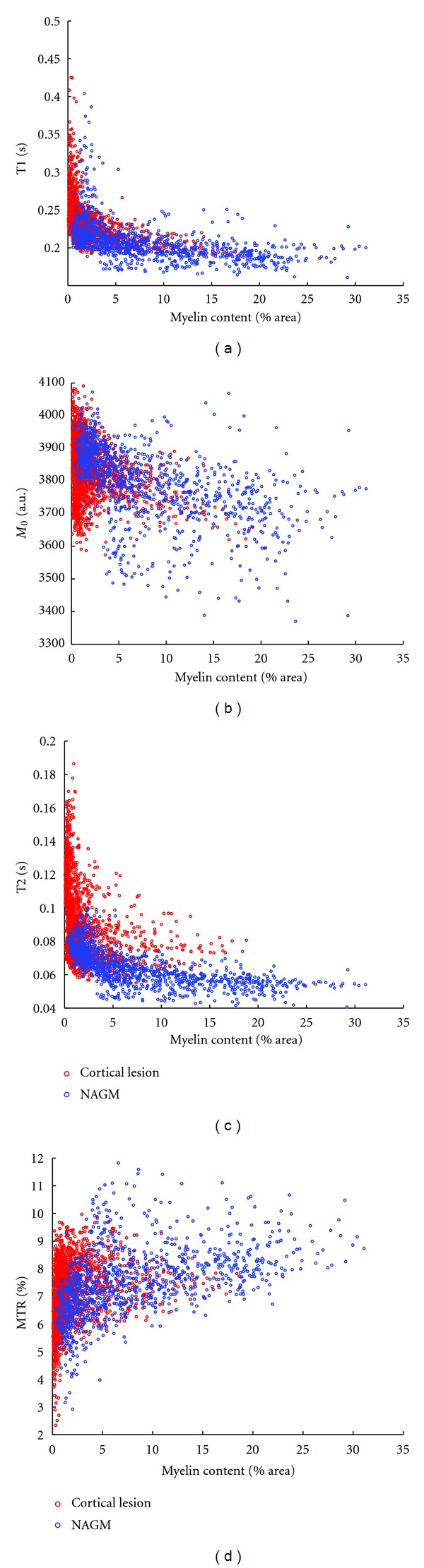
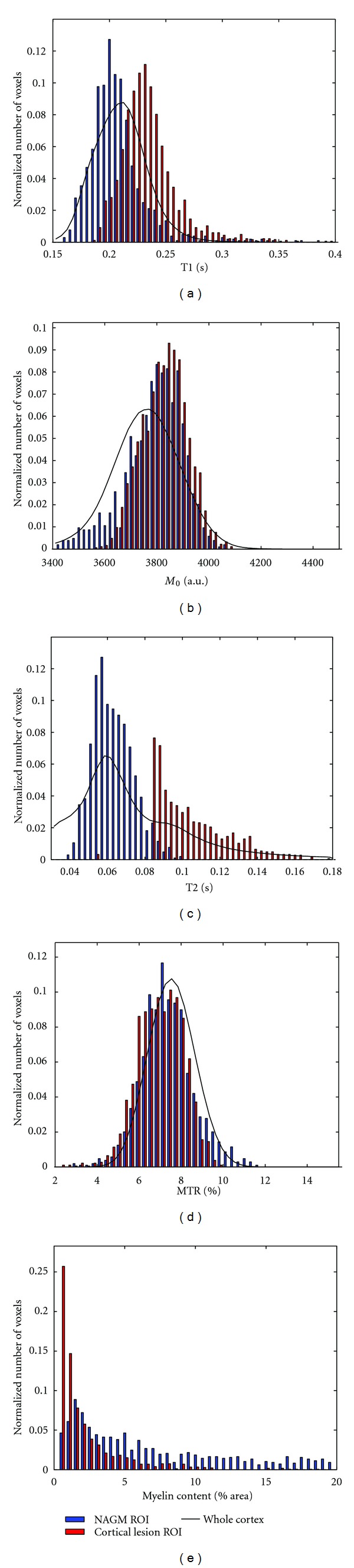
Although significant improvements have been made regarding the visualization and characterization of cortical multiple sclerosis (MS) lesions using magnetic resonance imaging (MRI), cortical lesions (CL) continue to be under-detected in vivo, and we have a limited understanding of the causes of GM pathology. The objective of this study was to characterize the MRI signature of CLs to help interpret the changes seen in vivo and elucidate the factors limiting their visualization. A quantitative 3D high-resolution (350 μm isotropic) MRI study at 3 Tesla of a fixed post mortem cerebral hemisphere from a patient with MS is presented in combination with matched immunohistochemistry. Type III subpial lesions are characterized by an increase in T1, T2 and M0, and a decrease in MTR in comparison to the normal appearing cortex (NAC). All quantitative MR parameters were associated with cortical GM myelin content, while T1 showed the strongest correlation. The histogram analysis showed extensive overlap between CL and NAC for all MR parameters and myelin content. This is due to the poor contrast in myelin content between CL and NAC in comparison to the variability in myelo-architecture throughout the healthy cortex. This latter comparison is highlighted by the representation of T1 times on cortical surfaces at several laminar depths.

摘要

尽管在利用磁共振成像(MRI)对皮质多发性硬化(MS)病变进行可视化和特征描述方面已取得显著进展,但皮质病变(CL)在活体中仍未得到充分检测,而且我们对灰质病理学的病因了解有限。本研究的目的是描述CL的MRI特征,以帮助解释活体中观察到的变化,并阐明限制其可视化的因素。本文介绍了一项针对一名MS患者的固定死后大脑半球在3特斯拉下进行的定量三维高分辨率(各向同性350μm)MRI研究,并结合了匹配的免疫组织化学分析。与正常外观皮质(NAC)相比,III型软膜下病变的特征是T1、T2和M0增加,MTR降低。所有定量MR参数均与皮质灰质髓鞘含量相关,而T1显示出最强的相关性。直方图分析表明,CL和NAC在所有MR参数和髓鞘含量方面存在广泛重叠。这是因为与整个健康皮质中髓鞘结构的变异性相比,CL和NAC之间的髓鞘含量对比度较差。通过在几个层深的皮质表面上表示T1时间突出了后一种比较。

https://cdn.ncbi.nlm.nih.gov/pmc/blobs/7899/3506905/65202f0aa093/MSI2012-742018.009.jpg
https://cdn.ncbi.nlm.nih.gov/pmc/blobs/7899/3506905/e8068d98dd39/MSI2012-742018.001.jpg
https://cdn.ncbi.nlm.nih.gov/pmc/blobs/7899/3506905/38055f3983f1/MSI2012-742018.002.jpg
https://cdn.ncbi.nlm.nih.gov/pmc/blobs/7899/3506905/acd23b545533/MSI2012-742018.003.jpg
https://cdn.ncbi.nlm.nih.gov/pmc/blobs/7899/3506905/0120b915b0cd/MSI2012-742018.004.jpg
https://cdn.ncbi.nlm.nih.gov/pmc/blobs/7899/3506905/46a95dbb92b1/MSI2012-742018.005.jpg
https://cdn.ncbi.nlm.nih.gov/pmc/blobs/7899/3506905/81fc330139b9/MSI2012-742018.006.jpg
https://cdn.ncbi.nlm.nih.gov/pmc/blobs/7899/3506905/e43403fecfd7/MSI2012-742018.007.jpg
https://cdn.ncbi.nlm.nih.gov/pmc/blobs/7899/3506905/b30ba014e77f/MSI2012-742018.008.jpg
https://cdn.ncbi.nlm.nih.gov/pmc/blobs/7899/3506905/65202f0aa093/MSI2012-742018.009.jpg

相似文献

1
Quantitative magnetic resonance imaging of cortical multiple sclerosis pathology.皮质多发性硬化病变的定量磁共振成像
Mult Scler Int. 2012;2012:742018. doi: 10.1155/2012/742018. Epub 2012 Nov 18.
2
Ultra-high field MTR and qR2* differentiates subpial cortical lesions from normal-appearing gray matter in multiple sclerosis.超高场 MTR 和 qR2* 可区分多发性硬化症中的软脑膜皮质病变与正常外观的灰质。
Mult Scler. 2016 Sep;22(10):1306-14. doi: 10.1177/1352458515620499. Epub 2015 Dec 16.
3
A gradient in cortical pathology in multiple sclerosis by in vivo quantitative 7 T imaging.通过活体定量7T成像观察多发性硬化症皮质病理中的梯度变化。
Brain. 2015 Apr;138(Pt 4):932-45. doi: 10.1093/brain/awv011. Epub 2015 Feb 12.
4
In vivo characterization of cortical and white matter neuroaxonal pathology in early multiple sclerosis.早期多发性硬化症中皮质和白质神经轴突病变的体内特征分析
Brain. 2017 Nov 1;140(11):2912-2926. doi: 10.1093/brain/awx247.
5
High field (9.4 Tesla) magnetic resonance imaging of cortical grey matter lesions in multiple sclerosis.多发性硬化症皮质灰质病变的高磁场(9.4 特斯拉)磁共振成像。
Brain. 2010 Mar;133(Pt 3):858-67. doi: 10.1093/brain/awp335. Epub 2010 Jan 31.
6
Increased cortical grey matter lesion detection in multiple sclerosis with 7 T MRI: a post-mortem verification study.7T MRI 检测多发性硬化症皮质灰质病变的增加:一项死后验证研究。
Brain. 2016 May;139(Pt 5):1472-81. doi: 10.1093/brain/aww037. Epub 2016 Mar 8.
7
A clinically feasible 7-Tesla protocol for the identification of cortical lesions in Multiple Sclerosis.一种在临床上可行的 7 特斯拉协议,用于识别多发性硬化症中的皮质病变。
Eur Radiol. 2020 Aug;30(8):4586-4594. doi: 10.1007/s00330-020-06803-y. Epub 2020 Mar 24.
8
Comparison of Multiple Sclerosis Cortical Lesion Types Detected by Multicontrast 3T and 7T MRI.多对比度 3T 和 7T MRI 检测多发性硬化皮质病变类型的比较。
AJNR Am J Neuroradiol. 2019 Jul;40(7):1162-1169. doi: 10.3174/ajnr.A6099. Epub 2019 Jun 20.
9
The association between intra- and juxta-cortical pathology and cognitive impairment in multiple sclerosis by quantitative T* mapping at 7 T MRI.7T磁共振成像定量T*图谱研究多发性硬化症皮质内及皮质旁病变与认知障碍的关系
Neuroimage Clin. 2016 Nov 3;12:879-886. doi: 10.1016/j.nicl.2016.11.001. eCollection 2016.
10
In vivo imaging of cortical pathology in multiple sclerosis using ultra-high field MRI.利用超高场磁共振成像对多发性硬化症皮质病理进行活体成像。
Neurology. 2009 Sep 22;73(12):941-8. doi: 10.1212/WNL.0b013e3181b64bf7. Epub 2009 Jul 29.

引用本文的文献

1
Inferring laminar origins of MEG signals with optically pumped magnetometers (OPMs): A simulation study.利用光泵磁力仪(OPM)推断脑磁图(MEG)信号的层状起源:一项模拟研究。
Imaging Neurosci (Camb). 2025 Jan 2;3. doi: 10.1162/imag_a_00410. eCollection 2025.
2
Heterochronous laminar maturation in the human prefrontal cortex.人类前额叶皮质中的异时层状成熟
bioRxiv. 2025 Jan 30:2025.01.30.635751. doi: 10.1101/2025.01.30.635751.
3
Correlations between postmortem quantitative MRI parameters and demyelination, axonal loss and gliosis in multiple sclerosis: A systematic review and meta-analysis.

本文引用的文献

1
Postmortem verification of MS cortical lesion detection with 3D DIR.利用 3D DIR 对 MS 皮质病变进行死后验证。
Neurology. 2012 Jan 31;78(5):302-8. doi: 10.1212/WNL.0b013e31824528a0. Epub 2012 Jan 4.
2
Inflammatory cortical demyelination in early multiple sclerosis.早期多发性硬化症中的炎症性皮质脱髓鞘。
N Engl J Med. 2011 Dec 8;365(23):2188-97. doi: 10.1056/NEJMoa1100648.
3
Focal cortical lesion detection in multiple sclerosis: 3 Tesla DIR versus 7 Tesla FLASH-T2.多发性硬化症中的局灶性皮质病变检测:3T DIR 与 7T FLASH-T2。
多发性硬化症中死后定量MRI参数与脱髓鞘、轴突丢失和胶质细胞增生之间的相关性:一项系统评价和荟萃分析。
Brain Imaging Behav. 2025 Apr;19(2):323-335. doi: 10.1007/s11682-025-00971-5. Epub 2025 Jan 27.
4
Evolution of peripheral nerve changes in early multiple sclerosis-a longitudinal MR neurography study.早期多发性硬化症周围神经变化的演变——一项纵向磁共振神经成像研究
Front Neurol. 2024 May 3;15:1335408. doi: 10.3389/fneur.2024.1335408. eCollection 2024.
5
SSL-QALAS: Self-Supervised Learning for rapid multiparameter estimation in quantitative MRI using 3D-QALAS.SSL-QALAS:基于 3D-QALAS 的用于定量 MRI 中快速多参数估计的自监督学习。
Magn Reson Med. 2023 Nov;90(5):2019-2032. doi: 10.1002/mrm.29786. Epub 2023 Jul 6.
6
Quantitative MR Neurography in Multifocal Motor Neuropathy and Amyotrophic Lateral Sclerosis.多灶性运动神经病和肌萎缩侧索硬化症的定量磁共振神经成像
Diagnostics (Basel). 2023 Mar 25;13(7):1237. doi: 10.3390/diagnostics13071237.
7
Postmortem quantitative MRI disentangles histological lesion types in multiple sclerosis.死后定量 MRI 可区分多发性硬化症的组织学病变类型。
Brain Pathol. 2023 Nov;33(6):e13136. doi: 10.1111/bpa.13136. Epub 2022 Dec 8.
8
Quantitative myelin imaging with MRI and PET: an overview of techniques and their validation status.定量髓鞘成像的 MRI 和 PET:技术概述及其验证状态。
Brain. 2023 Apr 19;146(4):1243-1266. doi: 10.1093/brain/awac436.
9
Motion-corrected 3D-EPTI with efficient 4D navigator acquisition for fast and robust whole-brain quantitative imaging.采用高效 4D 导航采集的运动校正 3D-EPTI,用于快速、稳健的全脑定量成像。
Magn Reson Med. 2022 Sep;88(3):1112-1125. doi: 10.1002/mrm.29277. Epub 2022 Apr 28.
10
3D Echo Planar Time-resolved Imaging (3D-EPTI) for ultrafast multi-parametric quantitative MRI.用于超快多参数定量 MRI 的 3D 回波平面时间分辨成像(3D-EPTI)。
Neuroimage. 2022 Apr 15;250:118963. doi: 10.1016/j.neuroimage.2022.118963. Epub 2022 Feb 2.
J Magn Reson Imaging. 2012 Mar;35(3):537-42. doi: 10.1002/jmri.22847. Epub 2011 Nov 1.
4
Cortical diffusion-tensor imaging abnormalities in multiple sclerosis: a 3-year longitudinal study.多发性硬化症的皮质弥散张量成像异常:一项 3 年纵向研究。
Radiology. 2011 Dec;261(3):891-8. doi: 10.1148/radiol.11110195. Epub 2011 Oct 26.
5
T1 and proton density at 7 T in patients with multiple sclerosis: an initial study.多发性硬化症患者的 T1 和 7T 质子密度:初步研究。
Magn Reson Imaging. 2012 Jan;30(1):19-25. doi: 10.1016/j.mri.2011.07.018. Epub 2011 Sep 19.
6
Lesion detection at seven Tesla in multiple sclerosis using magnetisation prepared 3D-FLAIR and 3D-DIR.在 7T 磁共振使用磁化准备 3D-FLAIR 和 3D-DIR 检测多发性硬化病变。
Eur Radiol. 2012 Jan;22(1):221-31. doi: 10.1007/s00330-011-2242-z. Epub 2011 Aug 27.
7
A case of multiple sclerosis presenting with inflammatory cortical demyelination.一例以炎症性皮质脱髓鞘为表现的多发性硬化症。
Neurology. 2011 May 17;76(20):1705-10. doi: 10.1212/WNL.0b013e31821a44f1.
8
Imaging the tip of the iceberg: visualization of cortical lesions in multiple sclerosis.冰山一角:多发性硬化症的皮质病变可视化。
Mult Scler. 2011 Oct;17(10):1202-10. doi: 10.1177/1352458511406575. Epub 2011 May 11.
9
In vivo evidence of disseminated subpial T2* signal changes in multiple sclerosis at 7 T: a surface-based analysis.7T 下多发性硬化症脑表面下 T2* 信号弥散性改变的体内证据:基于表面的分析。
Neuroimage. 2011 Jul 1;57(1):55-62. doi: 10.1016/j.neuroimage.2011.04.009. Epub 2011 Apr 13.
10
Microstructural Parcellation of the Human Cerebral Cortex - From Brodmann's Post-Mortem Map to in vivo Mapping with High-Field Magnetic Resonance Imaging.人类大脑皮层的微观结构分区——从布罗德曼的尸检图谱到高场磁共振成像的活体图谱
Front Hum Neurosci. 2011 Feb 18;5:19. doi: 10.3389/fnhum.2011.00019. eCollection 2011.