• 文献检索
  • 文档翻译
  • 深度研究
  • 学术资讯
  • Suppr Zotero 插件Zotero 插件
  • 邀请有礼
  • 套餐&价格
  • 历史记录
应用&插件
Suppr Zotero 插件Zotero 插件浏览器插件Mac 客户端Windows 客户端微信小程序
定价
高级版会员购买积分包购买API积分包
服务
文献检索文档翻译深度研究API 文档MCP 服务
关于我们
关于 Suppr公司介绍联系我们用户协议隐私条款
关注我们

Suppr 超能文献

核心技术专利:CN118964589B侵权必究
粤ICP备2023148730 号-1Suppr @ 2026

文献检索

告别复杂PubMed语法,用中文像聊天一样搜索,搜遍4000万医学文献。AI智能推荐,让科研检索更轻松。

立即免费搜索

文件翻译

保留排版,准确专业,支持PDF/Word/PPT等文件格式,支持 12+语言互译。

免费翻译文档

深度研究

AI帮你快速写综述,25分钟生成高质量综述,智能提取关键信息,辅助科研写作。

立即免费体验

多发性硬化症中的局灶性皮质病变检测:3T DIR 与 7T FLASH-T2。

Focal cortical lesion detection in multiple sclerosis: 3 Tesla DIR versus 7 Tesla FLASH-T2.

机构信息

Beth Israel Deaconess Medical Center, Boston, Massachusetts, USA.

出版信息

J Magn Reson Imaging. 2012 Mar;35(3):537-42. doi: 10.1002/jmri.22847. Epub 2011 Nov 1.

DOI:10.1002/jmri.22847
PMID:22045554
原文链接:https://pmc.ncbi.nlm.nih.gov/articles/PMC4198329/
Abstract

PURPOSE

To evaluate the inter-rater agreement of cortical lesion detection using 7 Tesla (T) FLASH-T2 and 3T DIR sequences.

MATERIALS AND METHODS

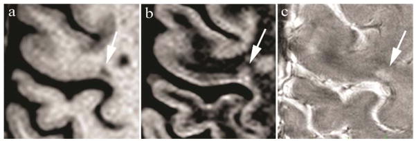
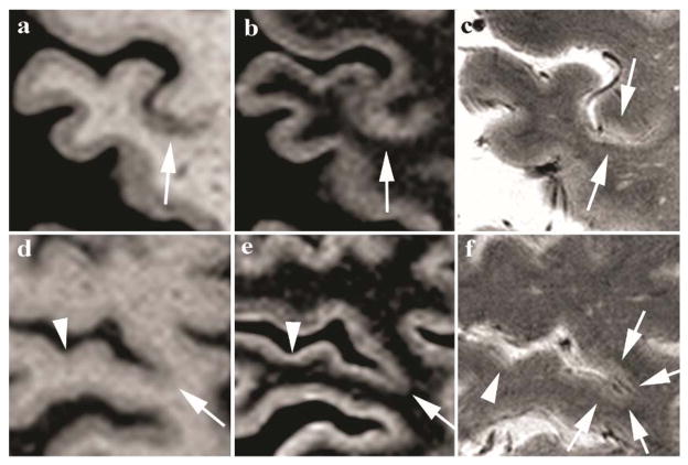
Twenty-six patients with multiple sclerosis were scanned on a human 7T (Siemens) and 3T MRI (TIM Trio, Siemens) to acquire 3T DIR/MEMPR and 7T FLASH-T2 sequences. Four independent reviewers scored and categorized cortical lesions in the bilateral precentral gyri (motor strips) as leukocortical, intracortical, or subpial. Inter-rater agreement was assessed according to lesion category using the kappa statistic. The sensitivity of recent MAGNIMS consensus guidelines for cortical lesion detection using 3T DIR was assessed with 7T FLASH-T2 as the reference gold standard.

RESULTS

Inter-rater agreement at 7T was excellent compared with 3T (k = 0.97 versus 0.12). FLASH-T2 at 7T detected subpial lesions while 3T DIR did not. The predicted sensitivity of 3T DIR sequence for cortical lesions in vivo is modest (range of 13.6 to 18.3%).

CONCLUSION

The 7T FLASH-T2 detects more cortical-particularly subpial-lesions compared with 3T DIR. In the absence of DIR/postmortem data, 7T FLASH-T2 is a suitable gold-standard instrument and should be incorporated into future consensus guidelines.

摘要

目的

评估使用 7 特斯拉(T)FLASH-T2 和 3T DIR 序列检测皮质病变的观察者间一致性。

材料与方法

26 例多发性硬化症患者在一台 7T(西门子)和 3T MRI(TIM Trio,西门子)上进行扫描,以获取 3T DIR/MEMPR 和 7T FLASH-T2 序列。四名独立的观察者对双侧中央前回(运动带)的皮质病变进行评分和分类,分为白质皮质、皮质内或皮质下病变。根据病变类别,使用kappa 统计评估观察者间的一致性。使用 7T FLASH-T2 作为参考金标准,评估 3T DIR 皮质病变检测的最新 MAGNIMS 共识指南的敏感性。

结果

与 3T 相比,7T 的观察者间一致性极好(k = 0.97 对 0.12)。FLASH-T2 在 7T 时可检测到皮质下病变,而 3T DIR 则不能。3T DIR 序列对皮质病变的预测敏感性适中(范围为 13.6%至 18.3%)。

结论

与 3T DIR 相比,7T FLASH-T2 可检测到更多的皮质病变,特别是皮质下病变。在缺乏 DIR/死后数据的情况下,7T FLASH-T2 是一种合适的金标准仪器,应纳入未来的共识指南。

https://cdn.ncbi.nlm.nih.gov/pmc/blobs/5fb5/4198329/343998579033/nihms632326f2.jpg
https://cdn.ncbi.nlm.nih.gov/pmc/blobs/5fb5/4198329/6c956e5023e0/nihms632326f1.jpg
https://cdn.ncbi.nlm.nih.gov/pmc/blobs/5fb5/4198329/343998579033/nihms632326f2.jpg
https://cdn.ncbi.nlm.nih.gov/pmc/blobs/5fb5/4198329/6c956e5023e0/nihms632326f1.jpg
https://cdn.ncbi.nlm.nih.gov/pmc/blobs/5fb5/4198329/343998579033/nihms632326f2.jpg

相似文献

1
Focal cortical lesion detection in multiple sclerosis: 3 Tesla DIR versus 7 Tesla FLASH-T2.多发性硬化症中的局灶性皮质病变检测:3T DIR 与 7T FLASH-T2。
J Magn Reson Imaging. 2012 Mar;35(3):537-42. doi: 10.1002/jmri.22847. Epub 2011 Nov 1.
2
Comparison of Multiple Sclerosis Cortical Lesion Types Detected by Multicontrast 3T and 7T MRI.多对比度 3T 和 7T MRI 检测多发性硬化皮质病变类型的比较。
AJNR Am J Neuroradiol. 2019 Jul;40(7):1162-1169. doi: 10.3174/ajnr.A6099. Epub 2019 Jun 20.
3
3 Tesla and 7 Tesla MRI of multiple sclerosis cortical lesions.3T 和 7T MRI 对多发性硬化皮质病变的研究。
J Magn Reson Imaging. 2010 Oct;32(4):971-7. doi: 10.1002/jmri.22115.
4
Improved detection of focal cortical lesions using 7T magnetisation transfer imaging in patients with multiple sclerosis.在多发性硬化症患者中使用7T磁化传递成像改善局灶性皮质病变的检测。
Mult Scler Relat Disord. 2014 Mar;3(2):258-65. doi: 10.1016/j.msard.2013.10.004. Epub 2013 Oct 19.
5
A comparison of 3T and 7T in the detection of small parenchymal veins within MS lesions.3T与7T在检测多发性硬化症病变内小实质静脉方面的比较。
Invest Radiol. 2009 Sep;44(9):491-4. doi: 10.1097/RLI.0b013e3181b4c144.
6
Inversion Recovery Susceptibility Weighted Imaging With Enhanced T2 Weighting at 3 T Improves Visualization of Subpial Cortical Multiple Sclerosis Lesions.3T 增强 T2 加权反转恢复敏感加权成像可提高脑皮层下多发性硬化病变的可视化效果。
Invest Radiol. 2020 Nov;55(11):727-735. doi: 10.1097/RLI.0000000000000698.
7
Lesion detection at seven Tesla in multiple sclerosis using magnetisation prepared 3D-FLAIR and 3D-DIR.在 7T 磁共振使用磁化准备 3D-FLAIR 和 3D-DIR 检测多发性硬化病变。
Eur Radiol. 2012 Jan;22(1):221-31. doi: 10.1007/s00330-011-2242-z. Epub 2011 Aug 27.
8
Double inversion recovery brain imaging at 3T: diagnostic value in the detection of multiple sclerosis lesions.3T下的双反转恢复脑成像:在检测多发性硬化症病变中的诊断价值
AJNR Am J Neuroradiol. 2007 Jan;28(1):54-9.
9
Seven-Tesla Magnetization Transfer Imaging to Detect Multiple Sclerosis White Matter Lesions.7特斯拉磁共振磁化传递成像检测多发性硬化症脑白质病变
J Neuroimaging. 2018 Mar;28(2):183-190. doi: 10.1111/jon.12474. Epub 2017 Sep 25.
10
Contribution of cortical lesion subtypes at 7T MRI to physical and cognitive performance in MS.7T MRI 下皮质病变亚型对 MS 患者躯体和认知功能的影响。
Neurology. 2013 Aug 13;81(7):641-9. doi: 10.1212/WNL.0b013e3182a08ce8. Epub 2013 Jul 17.

引用本文的文献

1
Fluid and White Matter Suppression contrasts MRI improves Deep Learning detection of Multiple Sclerosis Cortical Lesions.液体和白质抑制对比磁共振成像可改善深度学习对多发性硬化症皮质病变的检测。
Neuroimage Clin. 2025 Jul 14;48:103818. doi: 10.1016/j.nicl.2025.103818.
2
New Imaging Markers in Multiple Sclerosis and Related Disorders: Smoldering Inflammation and the Central Vein Sign.多发性硬化及相关疾病中的新型成像标志物:隐匿性炎症与中央静脉征
Neuroimaging Clin N Am. 2024 Aug;34(3):359-373. doi: 10.1016/j.nic.2024.03.004. Epub 2024 Apr 23.
3
Cortical lesions uniquely predict motor disability accrual and form rarely in the absence of new white matter lesions in multiple sclerosis.

本文引用的文献

1
Consensus recommendations for MS cortical lesion scoring using double inversion recovery MRI.多发性硬化症皮质病灶评分的双反转恢复 MRI 共识推荐意见。
Neurology. 2011 Feb 1;76(5):418-24. doi: 10.1212/WNL.0b013e31820a0cc4. Epub 2011 Jan 5.
2
3 Tesla and 7 Tesla MRI of multiple sclerosis cortical lesions.3T 和 7T MRI 对多发性硬化皮质病变的研究。
J Magn Reson Imaging. 2010 Oct;32(4):971-7. doi: 10.1002/jmri.22115.
3
Imaging cortical lesions in multiple sclerosis with ultra-high-field magnetic resonance imaging.利用超高场磁共振成像对多发性硬化症的皮质病变进行成像。
在多发性硬化症中,皮质病变独特地预示着运动功能障碍的累积,并且在没有新的白质病变的情况下很少出现。
medRxiv. 2023 Sep 25:2023.09.22.23295974. doi: 10.1101/2023.09.22.23295974.
4
Linking lesions in sensorimotor cortex to contralateral hand function in multiple sclerosis: a 7 T MRI study.多 sclerosis 中感觉运动皮层病灶与对侧手功能的关联:7 T MRI 研究。
Brain. 2022 Oct 21;145(10):3522-3535. doi: 10.1093/brain/awac203.
5
Multiple sclerosis cortical lesion detection with deep learning at ultra-high-field MRI.深度学习在超高场 MRI 下对多发性硬化皮质病变的检测。
NMR Biomed. 2022 Aug;35(8):e4730. doi: 10.1002/nbm.4730. Epub 2022 Mar 31.
6
Cortical lesion hotspots and association of subpial lesions with disability in multiple sclerosis.皮质病变热点与多发性硬化症软脑膜下病变相关性及其与残疾的关系。
Mult Scler. 2022 Aug;28(9):1351-1363. doi: 10.1177/13524585211069167. Epub 2022 Feb 10.
7
Cortical and phase rim lesions on 7 T MRI as markers of multiple sclerosis disease progression.7T磁共振成像上的皮质和相缘病变作为多发性硬化疾病进展的标志物
Brain Commun. 2021 Jun 24;3(3):fcab134. doi: 10.1093/braincomms/fcab134. eCollection 2021.
8
Imaging cortical multiple sclerosis lesions with ultra-high field MRI.超高场 MRI 成像皮质多发性硬化病变。
Neuroimage Clin. 2021;32:102847. doi: 10.1016/j.nicl.2021.102847. Epub 2021 Oct 6.
9
Sensitivity of T1/T2-weighted ratio in detection of cortical demyelination is similar to magnetization transfer ratio using post-mortem MRI.T1/T2 加权比检测皮质脱髓鞘的敏感性与使用死后 MRI 的磁化传递比相似。
Mult Scler. 2022 Feb;28(2):198-205. doi: 10.1177/13524585211014760. Epub 2021 May 20.
10
Prefrontal and hippocampal atrophy using 7-tesla magnetic resonance imaging in patients with Parkinson's disease.帕金森病患者使用7特斯拉磁共振成像检测前额叶和海马萎缩
Acta Radiol Open. 2021 Feb 24;10(2):2058460120988097. doi: 10.1177/2058460120988097. eCollection 2021 Feb.
Arch Neurol. 2010 Jul;67(7):812-8. doi: 10.1001/archneurol.2010.148.
4
In vivo imaging of cortical pathology in multiple sclerosis using ultra-high field MRI.利用超高场磁共振成像对多发性硬化症皮质病理进行活体成像。
Neurology. 2009 Sep 22;73(12):941-8. doi: 10.1212/WNL.0b013e3181b64bf7. Epub 2009 Jul 29.
5
Cognitive impairment in multiple sclerosis.多发性硬化症中的认知障碍。
Lancet Neurol. 2008 Dec;7(12):1139-51. doi: 10.1016/S1474-4422(08)70259-X.
6
3D MPRAGE improves classification of cortical lesions in multiple sclerosis.3D MPRAGE可改善多发性硬化症中皮质病变的分类。
Mult Scler. 2008 Nov;14(9):1214-9. doi: 10.1177/1352458508094644.
7
Multi-contrast, isotropic, single-slab 3D MR imaging in multiple sclerosis.多发性硬化症的多对比、各向同性、单层3D磁共振成像
Eur Radiol. 2008 Oct;18(10):2311-20. doi: 10.1007/s00330-008-1009-7. Epub 2008 May 29.
8
Brain morphometry with multiecho MPRAGE.采用多回波MPRAGE序列的脑形态测量
Neuroimage. 2008 Apr 1;40(2):559-569. doi: 10.1016/j.neuroimage.2007.12.025. Epub 2008 Feb 1.
9
Detection of cortical lesions is dependent on choice of slice thickness in patients with multiple sclerosis.在多发性硬化症患者中,皮质病变的检测取决于切片厚度的选择。
Int Rev Neurobiol. 2007;79:475-89. doi: 10.1016/S0074-7742(07)79021-9.
10
Extensive cortical remyelination in patients with chronic multiple sclerosis.慢性多发性硬化症患者广泛的皮质髓鞘再生
Brain Pathol. 2007 Apr;17(2):129-38. doi: 10.1111/j.1750-3639.2006.00043.x.