The University of Queensland, NHMRC Centre of Clinical Research Excellence in Spinal Pain, Injury and Health and School of Health and Rehabilitations Sciences, Brisbane, Queensland, Australia.
PLoS One. 2012;7(12):e51298. doi: 10.1371/journal.pone.0051298. Epub 2012 Dec 5.
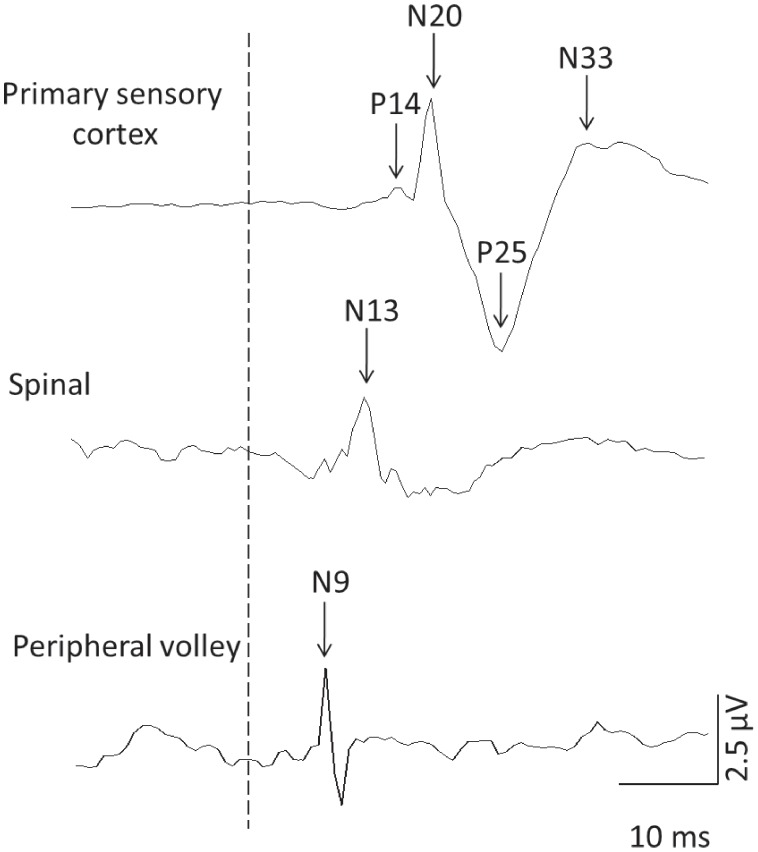
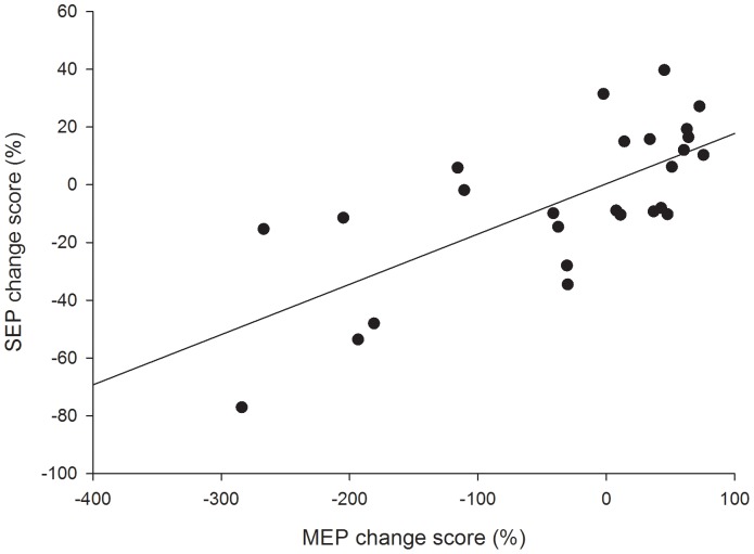
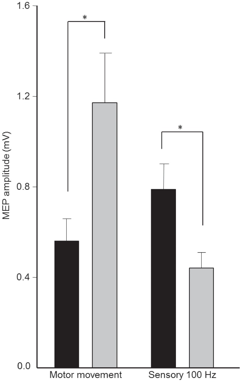
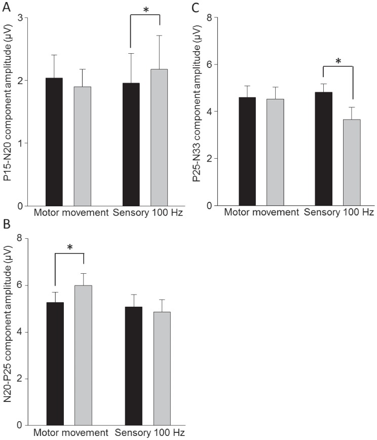
Peripheral electrical stimulation (PES) is a common clinical technique known to induce changes in corticomotor excitability; PES applied to induce a tetanic motor contraction increases, and PES at sub-motor threshold (sensory) intensities decreases, corticomotor excitability. Understanding of the mechanisms underlying these opposite changes in corticomotor excitability remains elusive. Modulation of primary sensory cortex (S1) excitability could underlie altered corticomotor excitability with PES. Here we examined whether changes in primary sensory (S1) and motor (M1) cortex excitability follow the same time-course when PES is applied using identical stimulus parameters. Corticomotor excitability was measured using transcranial magnetic stimulation (TMS) and sensory cortex excitability using somatosensory evoked potentials (SEPs) before and after 30 min of PES to right abductor pollicis brevis (APB). Two PES paradigms were tested in separate sessions; PES sufficient to induce a tetanic motor contraction (30-50 Hz; strong motor intensity) and PES at sub motor-threshold intensity (100 Hz). PES applied to induce strong activation of APB increased the size of the N(20)-P(25) component, thought to reflect sensory processing at cortical level, and increased corticomotor excitability. PES at sensory intensity decreased the size of the P25-N33 component and reduced corticomotor excitability. A positive correlation was observed between the changes in amplitude of the cortical SEP components and corticomotor excitability following sensory and motor PES. Sensory PES also increased the sub-cortical P(14)-N(20) SEP component. These findings provide evidence that PES results in co-modulation of S1 and M1 excitability, possibly due to cortico-cortical projections between S1 and M1. This mechanism may underpin changes in corticomotor excitability in response to afferent input generated by PES.
外周电刺激(PES)是一种常见的临床技术,已知其可引起运动皮质兴奋性的变化;应用于诱导强直运动收缩的 PES 增加,而应用于亚运动阈值(感觉)强度的 PES 则降低运动皮质兴奋性。对于这些运动皮质兴奋性相反变化的潜在机制仍然难以捉摸。初级感觉皮层(S1)兴奋性的调节可能是 PES 改变运动皮质兴奋性的基础。在这里,我们研究了当使用相同的刺激参数应用 PES 时,初级感觉(S1)和运动(M1)皮层兴奋性是否遵循相同的时间过程。使用经颅磁刺激(TMS)测量运动皮质兴奋性,使用体感诱发电位(SEP)测量感觉皮层兴奋性,在 30 分钟的 PES 前后对右侧外展拇指短肌(APB)进行测量。在单独的会话中测试了两种 PES 范式;足以引起强直运动收缩的 PES(30-50 Hz;强运动强度)和低于运动阈值强度的 PES(100 Hz)。应用于诱导 APB 强烈激活的 PES 增加了 N(20)-P(25)成分的大小,该成分被认为反映了皮质水平的感觉处理,并增加了运动皮质兴奋性。感觉强度的 PES 减小了 P25-N33 成分的大小,并降低了运动皮质兴奋性。在感觉和运动 PES 后,观察到皮质 SEP 成分的振幅变化与运动皮质兴奋性之间存在正相关。感觉 PES 还增加了皮质下的 P(14)-N(20)SEP 成分。这些发现提供了证据,表明 PES 导致 S1 和 M1 兴奋性的共同调节,这可能是由于 S1 和 M1 之间的皮质-皮质投射。该机制可能是 PES 产生的传入输入引起的运动皮质兴奋性变化的基础。