Institute of Liver and Biliary Sciences (ILBS), New Delhi, India.
Clin Transl Gastroenterol. 2012 Oct 4;3(10):e23. doi: 10.1038/ctg.2012.17.
CD4+ regulatory T cells (Tregs) seem to have a key role in persistence of hepatitis B virus (HBV) infection. Notch and transforming growth factor (TGF-β) signaling independently help in the differentiation and regulation of CD4+T cells, including T-helper (T(H)) 1, T(H)2, and Tregs. Whether, the two pathways have modulatory role on different stages of HBV infection and severity of liver disease is not clear. We investigated Notch and TGF-β families' gene expression in peripheral blood and intrahepatic lymphocytes in patients with different stages of chronic HBV (CHB) infection.
Peripheral blood mononuclear cells (PBMCs), CD4(+), and CD8(+) T cells were isolated from patients with acute HBV (AVH-B, n=15), CHB (n=16), and controls (HC, n=10). In addition to PBMCs, intrahepatic lymphocytes were obtained from liver biopsies from CHB (n=12), cirrhosis (n=12), hepatocellular carcinoma (HCC, n=5), and healthy livers (n=5). Notch family (Notch1-4, Hes1, Jag1, and NF-kβ) and TGF-β family gene expressions were studied by real-time PCR, flow cytometry, and immunohistochemistry.
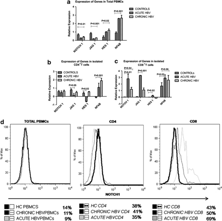
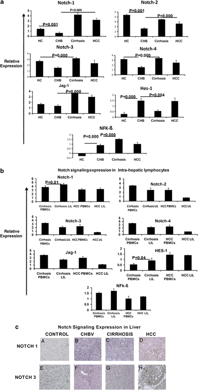
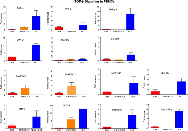
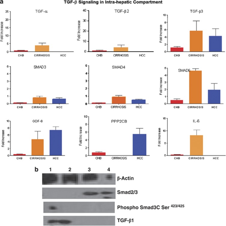
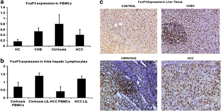
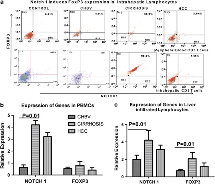
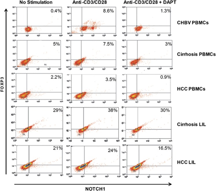
Relative expression of Notch signaling target genes, Hes1 and NF-kβ, was higher in the total PBMCs of AVH-B and CHB patients than that in HC patients (Log relative quantification (RQ); 1.1 AVH-B vs. 0.3 HC, 1.3 CHB vs. 0.3 HC; P=0.02). CD8(+) T cells showed upregulated expression of Hes1 and Notch1 (P=0.02 and 0.01, respectively) in AVH-B than in CHB patients. Also, in AVH-B patients, HBV-specific CD8(+) T-cell proliferation (5.74% vs. 2.7%) and TGF-β signaling activity were higher. All Notch receptors and ligands were upregulated in the PBMCs in CHB infection (CHB vs. cirrhosis, P=0.001; CHB vs. HCC, P=0.023; and cirrhosis vs. HCC, P=NS). Intrahepatic expression of Notch1 and FoxP3 were significantly higher in cirrhotics and HCCs, and further blockage of Notch signaling reduced the FoxP3 expression. Array data of TGF-β family showed increased TGF-β3, TGF-α, SMAD3, SMAD4, SMAD6, and GDF9 expression on intrahepatic lymphocytes in cirrhotic and HCC patients compared with CHB.
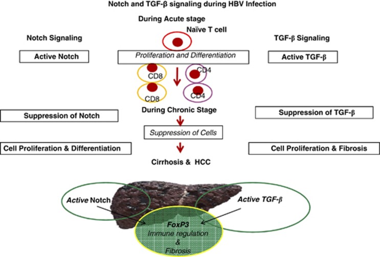
Our findings suggest that there is a complementary association between Notch1 and Hes1 in CD8(+)T cells during AVH-B infection. On development of CHB infection, repression of the Notch receptors mediates the regulation of immune response in patients, who progress to cirrhosis and HCC. Finally, HBV infection drives increased Notch1, TGF-β, and FoxP3 expression on intrahepatic T cells in cirrhosis, resulting in fibrogenesis and disease progression.
CD4+ 调节性 T 细胞(Tregs)似乎在乙型肝炎病毒(HBV)感染的持续存在中发挥关键作用。Notch 和转化生长因子(TGF-β)信号通路独立地有助于 CD4+T 细胞的分化和调节,包括辅助性 T(Th)1、Th2 和 Tregs。这两个途径是否对 HBV 感染的不同阶段和肝脏疾病的严重程度具有调节作用尚不清楚。我们研究了慢性乙型肝炎(CHB)感染不同阶段患者外周血和肝内淋巴细胞中 Notch 和 TGF-β 家族基因的表达。
从急性 HBV(AVH-B,n=15)、CHB(n=16)和对照组(HC,n=10)患者中分离外周血单核细胞(PBMCs)、CD4+和 CD8+T 细胞。除 PBMCs 外,还从 CHB(n=12)、肝硬化(n=12)、肝细胞癌(HCC,n=5)和健康肝脏(n=5)的肝活检中获得肝内淋巴细胞。通过实时 PCR、流式细胞术和免疫组织化学研究 Notch 家族(Notch1-4、Hes1、Jag1 和 NF-kβ)和 TGF-β 家族基因的表达。
AVH-B 和 CHB 患者的总 PBMCs 中 Notch 信号靶基因 Hes1 和 NF-kβ的相对表达量高于 HC 患者(Log 相对定量(RQ);1.1 AVH-B 比 0.3 HC,1.3 CHB 比 0.3 HC;P=0.02)。与 CHB 患者相比,AVH-B 患者的 CD8+T 细胞中 Hes1 和 Notch1 的表达上调(P=0.02 和 0.01)。此外,在 AVH-B 患者中,HBV 特异性 CD8+T 细胞增殖(5.74%比 2.7%)和 TGF-β 信号活性更高。CHB 感染时,所有 Notch 受体和配体在 PBMCs 中均上调(CHB 与肝硬化,P=0.001;CHB 与 HCC,P=0.023;肝硬化与 HCC,P=NS)。在肝硬化和 HCC 患者中,肝内 Notch1 和 FoxP3 的表达明显升高,进一步阻断 Notch 信号可降低 FoxP3 的表达。TGF-β 家族的阵列数据显示,与 CHB 相比,肝硬化和 HCC 患者肝内淋巴细胞中 TGF-β3、TGF-α、SMAD3、SMAD4、SMAD6 和 GDF9 的表达增加。
我们的研究结果表明,在 AVH-B 感染期间,CD8+T 细胞中存在 Notch1 和 Hes1 之间的互补关联。在 CHB 感染发展过程中,Notch 受体的抑制调节了进展为肝硬化和 HCC 的患者的免疫反应。最后,HBV 感染导致肝硬化患者肝内 T 细胞中 Notch1、TGF-β 和 FoxP3 的表达增加,导致纤维化和疾病进展。