Hormones and Cancer Division, Kolling Institute of Medical Research, University of Sydney, Royal North Shore Hospital, St Leonards, New South Wales, Australia.
Br J Cancer. 2013 Feb 5;108(2):351-60. doi: 10.1038/bjc.2012.552. Epub 2013 Jan 8.
Tissue protein expression profiling has the potential to detect new biomarkers to improve breast cancer (BC) diagnosis, staging, and prognostication. This study aimed to identify tissue proteins that differentiate breast cancer tissue from healthy breast tissue using protein chip mass spectrometry and to examine associations with conventional pathological features.
To develop a training model, 82 BC and 82 adjacent unaffected tissue (AT) samples were analysed on cation-exchange protein chips by time-of-flight mass spectrometry. For validation, 89 independent BC and AT sample pairs were analysed.
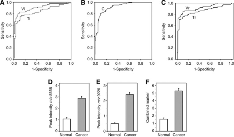
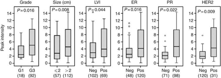
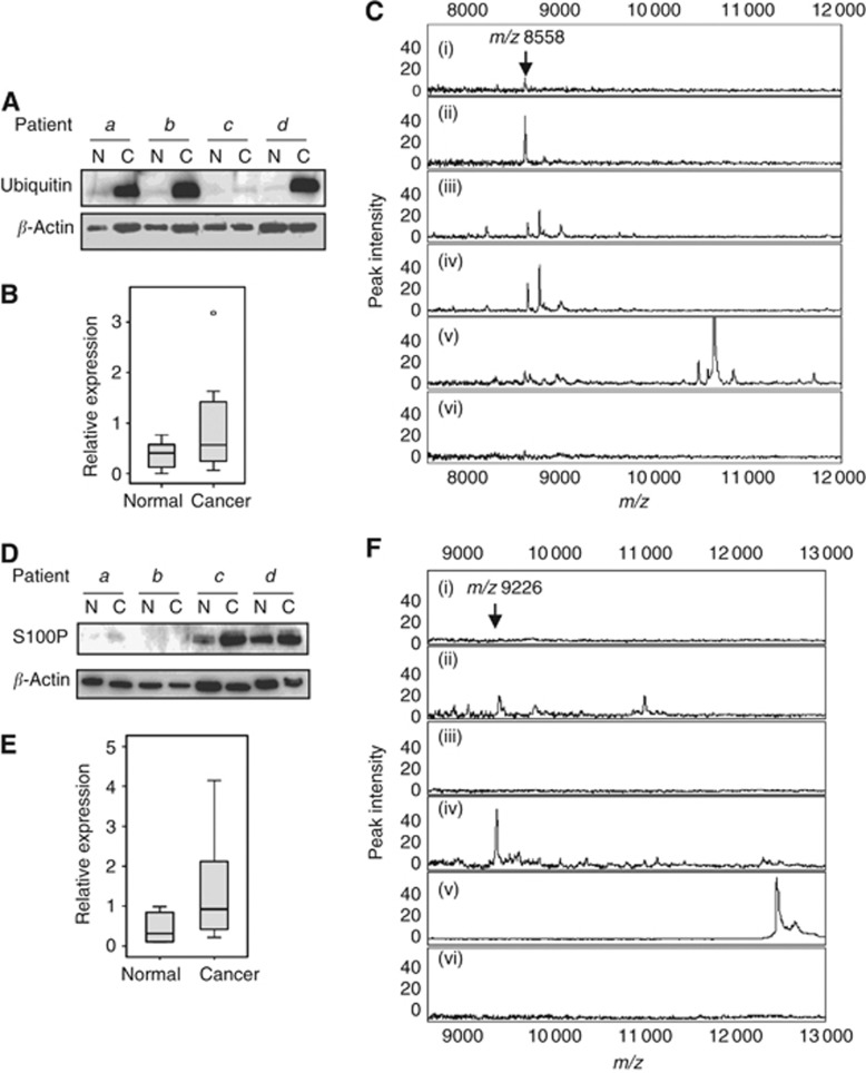
From the protein peaks that were differentially expressed between BC and AT by univariate analysis, binary logistic regression yielded two peaks that together classified BC and AT with a ROC area under the curve of 0.92. Two proteins, ubiquitin and S100P (in a novel truncated form), were identified by liquid chromatography/tandem mass spectrometry and validated by immunoblotting and reactive-surface protein chip immunocapture. The combined marker panel was positively associated with high histologic grade, larger tumour size, lymphovascular invasion, ER and PR positivity, and HER2 overexpression, suggesting that it may be associated with a HER2-enriched molecular subtype of breast cancer.
This independently validated protein panel may be valuable in the classification and prognostication of breast cancer patients.
组织蛋白表达谱分析有可能发现新的生物标志物,从而改善乳腺癌(BC)的诊断、分期和预后判断。本研究旨在利用蛋白芯片质谱技术鉴定区分乳腺癌组织和健康乳腺组织的组织蛋白,并探讨其与常规病理特征的关联。
为了建立训练模型,我们采用飞行时间质谱技术对 82 例乳腺癌和 82 例相邻无病变组织(AT)样本进行阳离子交换蛋白芯片分析。为了验证,我们对 89 对独立的 BC 和 AT 样本进行了分析。
通过单变量分析,从 BC 和 AT 之间差异表达的蛋白峰中,二元逻辑回归得到了两个峰,它们共同将 BC 和 AT 分类,ROC 曲线下面积为 0.92。通过液相色谱/串联质谱鉴定出两种蛋白,泛素和 S100P(一种新的截断形式),并用免疫印迹和反应表面蛋白芯片免疫捕获进行了验证。联合标志物组合与高组织学分级、更大的肿瘤大小、淋巴血管侵犯、ER 和 PR 阳性以及 HER2 过表达呈正相关,提示其可能与富含 HER2 的乳腺癌分子亚型有关。
这个独立验证的蛋白谱可能对乳腺癌患者的分类和预后判断具有重要价值。