• 文献检索
  • 文档翻译
  • 深度研究
  • 学术资讯
  • Suppr Zotero 插件Zotero 插件
  • 邀请有礼
  • 套餐&价格
  • 历史记录
应用&插件
Suppr Zotero 插件Zotero 插件浏览器插件Mac 客户端Windows 客户端微信小程序
定价
高级版会员购买积分包购买API积分包
服务
文献检索文档翻译深度研究API 文档MCP 服务
关于我们
关于 Suppr公司介绍联系我们用户协议隐私条款
关注我们

Suppr 超能文献

核心技术专利:CN118964589B侵权必究
粤ICP备2023148730 号-1Suppr @ 2026

文献检索

告别复杂PubMed语法,用中文像聊天一样搜索,搜遍4000万医学文献。AI智能推荐,让科研检索更轻松。

立即免费搜索

文件翻译

保留排版,准确专业,支持PDF/Word/PPT等文件格式,支持 12+语言互译。

免费翻译文档

深度研究

AI帮你快速写综述,25分钟生成高质量综述,智能提取关键信息,辅助科研写作。

立即免费体验

使用切伦科夫激发荧光的光子束投影成像。

Projection imaging of photon beams using Čerenkov-excited fluorescence.

机构信息

Thayer School of Engineering, Dartmouth College, Hanover, NH 03755, USA.

出版信息

Phys Med Biol. 2013 Feb 7;58(3):601-19. doi: 10.1088/0031-9155/58/3/601. Epub 2013 Jan 14.

DOI:10.1088/0031-9155/58/3/601
PMID:23318469
原文链接:https://pmc.ncbi.nlm.nih.gov/articles/PMC3568662/
Abstract

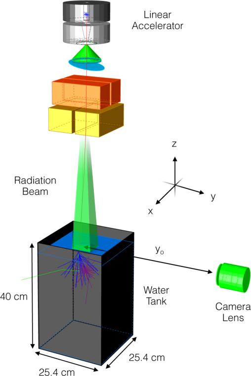
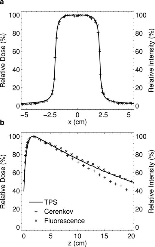
Full 3D beam profiling and quality assurance (QA) of therapeutic megavoltage linear accelerator (LINAC) x-ray photon beams is not routinely performed due to the slow point-by-point measurement nature of conventional scanning ionization chamber systems. In this study we explore a novel optical-based dose imaging approach using a standard commercial camera, water tank, and fluorescent dye, which when excited by the Čerenkov emission induced by the radiation beam, allows 2D projection imaging in a fast timeframe, potentially leading toward 3D tomographic beam profiling. Detailed analysis was carried out to optimize the imaging parameters in the experimental setup. The results demonstrate that the captured images are linear with delivered dose, independent of dose rate, and comparison of experimentally captured images to a reference dose distribution for a 4 × 4 cm(2) 6 MV x-ray photon beam yielded results with improved accuracy over a previous study which used direct imaging and Monte Carlo calibration of the Čerenkov emission itself. The agreement with the reference dose distribution was within 1-2% in the lateral direction, and ±3% in the depth direction. The study was restricted to single 2D image projection, with the eventual goal of creating full 3D profiles after tomographic reconstruction from multiple projections. Given the increasingly complex advances in radiation therapy, and the increased emphasis on patient-specific treatment plans, further refinement of the technique could prove to be an important tool for fast and robust QA of x-ray photon LINAC beams.

摘要

由于传统扫描电离室系统的逐点测量性质,对治疗兆伏级直线加速器 (LINAC) X 射线光子束进行全 3D 束流特性分析和质量保证 (QA) 通常无法实现。在这项研究中,我们探索了一种新颖的基于光学的剂量成像方法,使用标准的商业相机、水箱和荧光染料,当被辐射束产生的切伦科夫发射激发时,允许在快速时间框架内进行 2D 投影成像,从而有可能实现 3D 层析束流特性分析。我们进行了详细的分析以优化实验设置中的成像参数。结果表明,捕获的图像与所传递的剂量呈线性关系,与剂量率无关,并且将实验捕获的图像与 4×4cm(2)6MV X 射线光子束的参考剂量分布进行比较,结果的准确性优于之前使用直接成像和切伦科夫发射本身的蒙特卡罗校准的研究。在侧向方向上的误差在 1%到 2%之间,在深度方向上的误差在 ±3%之间。该研究仅限于单个 2D 图像投影,最终目标是在从多个投影进行层析重建后创建完整的 3D 轮廓。鉴于放射治疗的日益复杂以及对患者特异性治疗计划的重视,该技术的进一步改进可能成为 X 射线光子 LINAC 束快速和强大 QA 的重要工具。

https://cdn.ncbi.nlm.nih.gov/pmc/blobs/ee6d/3568662/b16777ec48d6/nihms-438713-f0009.jpg
https://cdn.ncbi.nlm.nih.gov/pmc/blobs/ee6d/3568662/6712fc54082d/nihms-438713-f0001.jpg
https://cdn.ncbi.nlm.nih.gov/pmc/blobs/ee6d/3568662/d966bb44e9fb/nihms-438713-f0002.jpg
https://cdn.ncbi.nlm.nih.gov/pmc/blobs/ee6d/3568662/8b41f32a352b/nihms-438713-f0003.jpg
https://cdn.ncbi.nlm.nih.gov/pmc/blobs/ee6d/3568662/a8a3c7d9ebac/nihms-438713-f0004.jpg
https://cdn.ncbi.nlm.nih.gov/pmc/blobs/ee6d/3568662/cc34c930d24e/nihms-438713-f0005.jpg
https://cdn.ncbi.nlm.nih.gov/pmc/blobs/ee6d/3568662/3940f1ad2dc6/nihms-438713-f0006.jpg
https://cdn.ncbi.nlm.nih.gov/pmc/blobs/ee6d/3568662/7641cadfc163/nihms-438713-f0007.jpg
https://cdn.ncbi.nlm.nih.gov/pmc/blobs/ee6d/3568662/a14581fed8ef/nihms-438713-f0008.jpg
https://cdn.ncbi.nlm.nih.gov/pmc/blobs/ee6d/3568662/b16777ec48d6/nihms-438713-f0009.jpg

相似文献

1
Projection imaging of photon beams using Čerenkov-excited fluorescence.使用切伦科夫激发荧光的光子束投影成像。
Phys Med Biol. 2013 Feb 7;58(3):601-19. doi: 10.1088/0031-9155/58/3/601. Epub 2013 Jan 14.
2
Projection imaging of photon beams by the Čerenkov effect.切伦科夫效应的光子束投影成像。
Med Phys. 2013 Jan;40(1):012101. doi: 10.1118/1.4770286.
3
Optical cone beam tomography of Cherenkov-mediated signals for fast 3D dosimetry of x-ray photon beams in water.用于水中X射线光子束快速三维剂量测定的切伦科夫介导信号的光学锥束断层扫描
Med Phys. 2015 Jul;42(7):4127-36. doi: 10.1118/1.4922135.
4
Investigation of the use of external aluminium targets for portal imaging in a medical accelerator using Geant4 Monte Carlo simulation.使用Geant4蒙特卡罗模拟研究医用加速器中用于射野成像的外部铝靶的应用。
Br J Radiol. 2018 Apr;91(1084):20170376. doi: 10.1259/bjr.20170376. Epub 2018 Feb 6.
5
Ion chamber and film-based quality assurance of mixed electron-photon radiation therapy.混合电子-光子放射治疗的离子室和膜基质量保证。
Med Phys. 2021 Sep;48(9):5382-5395. doi: 10.1002/mp.15081. Epub 2021 Jul 29.
6
Cherenkov imaging for linac beam shape analysis as a remote electronic quality assessment verification tool.切伦科夫成像在直线加速器束流形状分析中的应用,作为一种远程电子质量评估验证工具。
Med Phys. 2019 Feb;46(2):811-821. doi: 10.1002/mp.13303. Epub 2018 Dec 14.
7
Beam perturbation characteristics of a 2D transmission silicon diode array, Magic Plate.二维透射硅二极管阵列Magic Plate的射束扰动特性
J Appl Clin Med Phys. 2016 Mar 8;17(2):85-98. doi: 10.1120/jacmp.v17i2.5932.
8
Imaging Cerenkov emission as a quality assurance tool in electron radiotherapy.将切伦科夫辐射成像作为电子放射治疗中的质量保证工具。
Phys Med Biol. 2014 Apr 21;59(8):1963-78. doi: 10.1088/0031-9155/59/8/1963. Epub 2014 Apr 2.
9
A photon source model based on particle transport in a parameterized accelerator structure for Monte Carlo dose calculations.基于蒙特卡罗剂量计算中参数化加速器结构中粒子输运的光子源模型。
Med Phys. 2018 Jul;45(7):2937-2946. doi: 10.1002/mp.12976. Epub 2018 Jun 3.
10
Experimental verification of a commercial Monte Carlo-based dose calculation module for high-energy photon beams.高能光子束商用蒙特卡罗剂量计算模块的实验验证。
Phys Med Biol. 2009 Dec 21;54(24):7363-77. doi: 10.1088/0031-9155/54/24/008. Epub 2009 Nov 24.

引用本文的文献

1
Preliminary study of luminescence phenomena from various materials under ultra-high dose rate proton beam irradiation for dose management.超高剂量率质子束辐照下各种材料发光现象用于剂量管理的初步研究。
Sci Rep. 2024 Jun 24;14(1):14504. doi: 10.1038/s41598-024-65513-3.
2
Scintillation of polyester fabric and clothing via proton irradiation and its utilization in surface imaging of proton pencil beams.通过质子辐照实现聚酯织物和衣物的闪烁及其在质子笔形束表面成像中的应用。
Sci Rep. 2024 Jun 12;14(1):13494. doi: 10.1038/s41598-024-62456-7.
3
Direct in-water radiation dose measurements using Cherenkov emission corrected signals from polarization imaging for a clinical radiotherapy application.

本文引用的文献

1
Projection imaging of photon beams by the Čerenkov effect.切伦科夫效应的光子束投影成像。
Med Phys. 2013 Jan;40(1):012101. doi: 10.1118/1.4770286.
2
Time-gated Cherenkov emission spectroscopy from linear accelerator irradiation of tissue phantoms.组织体模的线性加速器辐照的时门控切伦科夫发射光谱学。
Opt Lett. 2012 Apr 1;37(7):1193-5. doi: 10.1364/OL.37.001193.
3
Verification of proton range, position, and intensity in IMPT with a 3D liquid scintillator detector system.利用 3D 液体闪烁探测器系统验证 IMPT 中的质子射程、位置和强度。
使用极化成像中的切伦科夫发射校正信号进行临床放射治疗中的直接水中辐射剂量测量。
Sci Rep. 2022 Jun 10;12(1):9608. doi: 10.1038/s41598-022-12672-w.
4
Survey of X-ray induced Cherenkov excited fluorophores with potential for human use.用于人体的 X 射线诱导切伦科夫辐射荧光团的调查。
J Radiat Res. 2021 Sep 13;62(5):833-840. doi: 10.1093/jrr/rrab055.
5
Increase in the intensity of an optical signal with fluorescein during proton and carbon-ion irradiation.荧光素在质子和碳离子辐照下增强了光学信号的强度。
J Appl Clin Med Phys. 2021 Jul;22(7):188-197. doi: 10.1002/acm2.13309. Epub 2021 Jun 14.
6
Optical imaging of muons.μ 子光学成像。
Sci Rep. 2020 Nov 26;10(1):20790. doi: 10.1038/s41598-020-76652-8.
7
Imaging Cherenkov emission for quality assurance of high-dose-rate brachytherapy.用于高剂量率近距离放射治疗质量保证的切伦科夫发射成像。
Sci Rep. 2020 Feb 27;10(1):3572. doi: 10.1038/s41598-020-60519-z.
8
Optical imaging provides rapid verification of static small beams, radiosurgery, and VMAT plans with millimeter resolution.光学成像是一种快速验证静态小射束、放射外科和 VMAT 计划的方法,其分辨率可达毫米级。
Med Phys. 2019 Nov;46(11):5227-5237. doi: 10.1002/mp.13797. Epub 2019 Oct 4.
9
Estimation of the fractions of luminescence of water at higher energy than Cerenkov-light threshold for various types of radiation.估算各种类型辐射的超过切伦科夫光阈值的水中的光的分数。
J Biomed Opt. 2019 Jun;24(6):1-9. doi: 10.1117/1.JBO.24.6.066005.
10
Radioluminescence in biomedicine: physics, applications, and models.生物医学中的放射发光:物理、应用和模型。
Phys Med Biol. 2019 Feb 6;64(4):04TR01. doi: 10.1088/1361-6560/aaf4de.
Med Phys. 2012 Mar;39(3):1239-46. doi: 10.1118/1.3681948.
4
A new water-equivalent 2D plastic scintillation detectors array for the dosimetry of megavoltage energy photon beams in radiation therapy.一种用于放射治疗中兆伏级能量光子束剂量学的新型水等效 2D 塑料闪烁体探测器阵列。
Med Phys. 2011 Dec;38(12):6763-74. doi: 10.1118/1.3664007.
5
Cerenkov emission induced by external beam radiation stimulates molecular fluorescence.外照射辐射引起的切伦科夫辐射会刺激分子荧光。
Med Phys. 2011 Jul;38(7):4127-32. doi: 10.1118/1.3592646.
6
Plastic scintillation dosimetry: comparison of three solutions for the Cerenkov challenge.塑料闪烁体剂量学:三种解决方案用于切伦科夫挑战的比较。
Phys Med Biol. 2011 Sep 21;56(18):5805-21. doi: 10.1088/0031-9155/56/18/003. Epub 2011 Aug 16.
7
Technical note: removing the stem effect when performing Ir-192 HDR brachytherapy in vivo dosimetry using plastic scintillation detectors: a relevant and necessary step.技术说明:在使用塑料闪烁探测器进行 Ir-192 HDR 近距离治疗体内剂量学测量时消除茎效应:一个相关且必要的步骤。
Med Phys. 2011 Apr;38(4):2176-9. doi: 10.1118/1.3562902.
8
Cerenkov radiation energy transfer (CRET) imaging: a novel method for optical imaging of PET isotopes in biological systems.切伦科夫辐射能量转移(CRET)成像:一种用于生物系统中 PET 同位素光学成像的新方法。
PLoS One. 2010 Oct 11;5(10):e13300. doi: 10.1371/journal.pone.0013300.
9
Exploration of the potential of liquid scintillators for real-time 3D dosimetry of intensity modulated proton beams.液体闪烁体用于调强质子束实时三维剂量测定潜力的探索。
Med Phys. 2009 May;36(5):1736-43. doi: 10.1118/1.3117583.
10
Liquid scintillator for 2D dosimetry for high-energy photon beams.用于高能光子束二维剂量测定的液体闪烁体。
Med Phys. 2009 May;36(5):1478-85. doi: 10.1118/1.3106390.