Choudhary Priyanka, Sudhamani S, Pandit Ajita, Kiri Vm
Department of Pathology, Dr. D.Y. Patil Medical College, Hospital and Research Center, Nerul, Navi Mumbai, India.
J Cytol. 2012 Oct;29(4):241-5. doi: 10.4103/0970-9371.103942.
Since the first introduction of Papanicolaou (Pap) stain in 1942 there have been many modifications. Of these, the Ultra-Fast Pap stain has become popular. This technique was further modified in India as many of the reagents were not available in our country. Our study was conducted by adapting this modified staining technique which involves the replacement of Gill's hematoxylin with Harris hematoxylin.
The aim of our prospective study was to assess the use of the modified Ultra-Fast Pap stain (MUFP) for fine needle aspiration cytology (FNAC) of various organs in comparison with the standard rapid Pap stain.
A total of 100 FNAC cases were studied by random sampling. Two smears were prepared for each case and stained by both, the MUFP and the rapid Pap stain. Scores were given and the quality index was calculated, followed by the statistical analysis. The number of cases was as follows: lymph node (43), thyroid (25), breast (23), salivary gland (02), and soft tissues (07). Scores were given on four parameters: Background of smears, overall staining pattern, cell morphology and nuclear staining. Quality index was calculated from the ratio of score achieved to the maximum score possible.
Results were analyzed using Mean, Median, Standard Deviation, 't' paired test, 'P' value and M-diff for statistical significance.
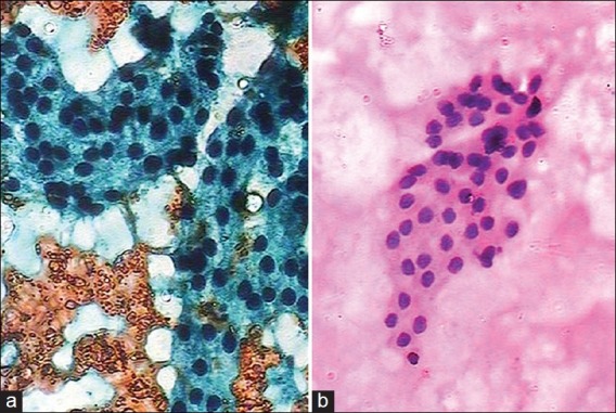
Correct diagnosis was made in all cases. The quality index of MUFP smears was better compared to the rapid Pap stain in all the organs, and was statistically significant. MUFP smears showed a clear red blood cells background, transparent cytoplasm and crisp nuclear features.
MUFP is a reliable and rapid technique for cytology diagnosis.
自1942年首次引入巴氏染色法以来,该方法已经历了多次改进。其中,超快速巴氏染色法已广泛应用。由于我国无法获取许多试剂,该技术在印度得到了进一步改进。我们的研究采用了这种改进的染色技术,即用哈里斯苏木精替代吉尔苏木精。
我们前瞻性研究的目的是评估改良超快速巴氏染色法(MUFP)在各器官细针穿刺细胞学检查(FNAC)中的应用,并与标准快速巴氏染色法进行比较。
通过随机抽样对100例FNAC病例进行研究。为每个病例制备两张涂片,分别用MUFP和快速巴氏染色法染色。给出评分并计算质量指数,随后进行统计分析。病例数量如下:淋巴结(43例)、甲状腺(25例)、乳腺(23例)、唾液腺(2例)和软组织(7例)。从涂片背景、整体染色模式、细胞形态和核染色四个参数进行评分。质量指数通过获得的分数与可能的最高分的比值计算得出。
使用均值、中位数、标准差、配对t检验、P值和M差异分析结果的统计学意义。
所有病例均做出了正确诊断。在所有器官中,MUFP涂片的质量指数均优于快速巴氏染色法,且具有统计学意义。MUFP涂片显示出清晰的红细胞背景、透明的细胞质和清晰的核特征。
MUFP是一种可靠且快速的细胞学诊断技术。