Department of Chemistry, Duke University, Durham, North Carolina 27708, USA.
J Invest Dermatol. 2013 Jul;133(7):1822-6. doi: 10.1038/jid.2013.37. Epub 2013 Jan 25.
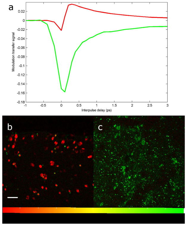
Pump-probe microscopy nondestructively differentiates eumelanin and pheomelanin and can be used to quantify melanin distributions in thin biopsy slices. Here we have extended that work for imaging eumelanin and pheomelanin distributions on a subcellular scale, allowing elucidation of characteristics of different cell types. The results show that melanin heterogeneity, previously found to be characteristic of melanomas, persists on the subcellular scale. We have also found spectral changes associated with melanin located in melanophages that could potentially differentiate invasive pigmented melanocytes from melanophages without immunohistochemical staining.
泵浦探针显微镜可无损地区分真黑色素和褐黑色素,并可用于定量分析薄活检切片中的黑色素分布。在这里,我们将该技术扩展到亚细胞尺度,以成像真黑色素和褐黑色素的分布,从而能够阐明不同细胞类型的特征。结果表明,先前发现黑色素瘤特有的黑色素异质性在亚细胞尺度上仍然存在。我们还发现了与位于黑色素细胞中的黑色素相关的光谱变化,这些变化可能无需免疫组织化学染色即可将侵袭性色素性黑色素细胞与黑色素细胞区分开来。