Beeman Scott C, Mandarino Lawrence J, Georges Joseph F, Bennett Kevin M
Department of Structural Biology, St. Jude Children's Research Hospital, Memphis, Tennessee.
Magn Reson Med. 2013 Dec;70(6):1728-38. doi: 10.1002/mrm.24619.
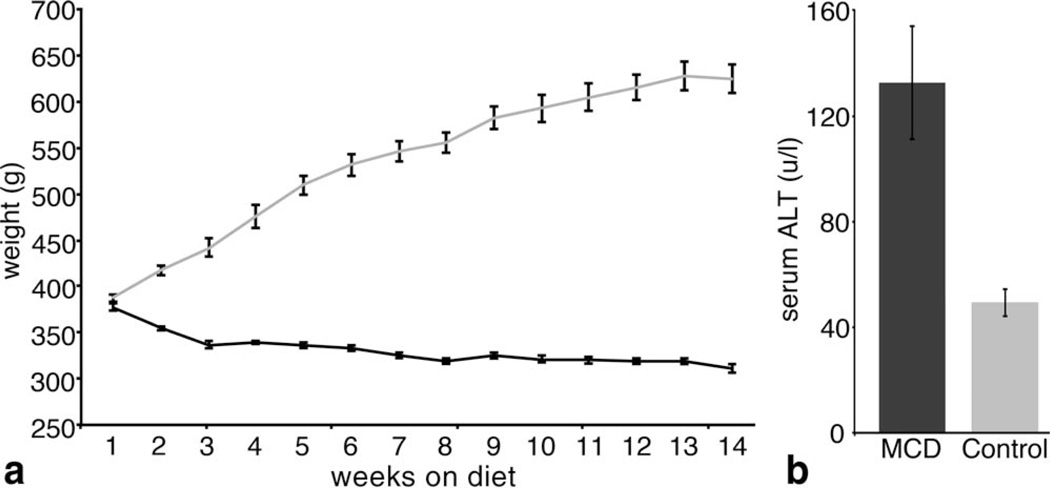
The goal of this work was to detect disease-related microstructural changes to the liver using magnetic resonance imaging. Chronic liver disease can cause microstructural changes in the liver that reduce plasma access to the perisinusoidal space--the site of exchange between the blood plasma and the hepatic parenchyma. The reduced plasma access to the perisinusoidal space inhibits hepatic function and contributes to the ∼30,000 chronic liver disease-related deaths per year.
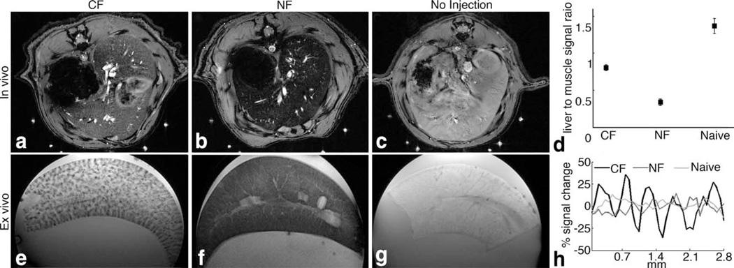
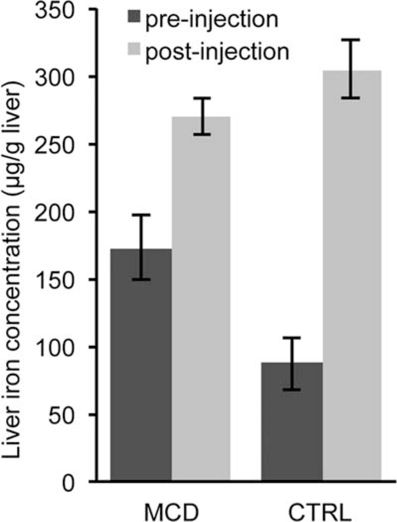
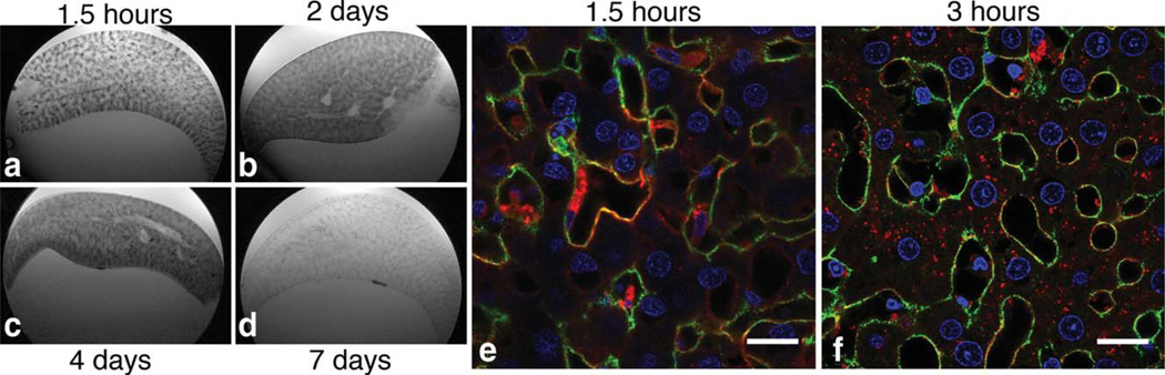
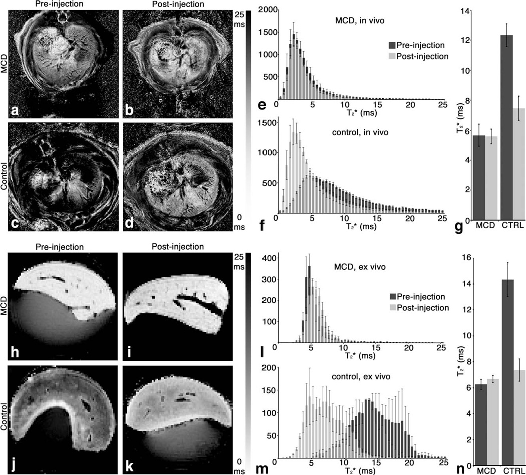
The extracellular matrix-specific cationized ferritin magnetic resonance imaging probe was injected intravenously into healthy rats and a rat model of the chronic liver disease non-alcoholic steatohepatitis. Rats were subsequently imaged with T2*-weighted magnetic resonance imaging.
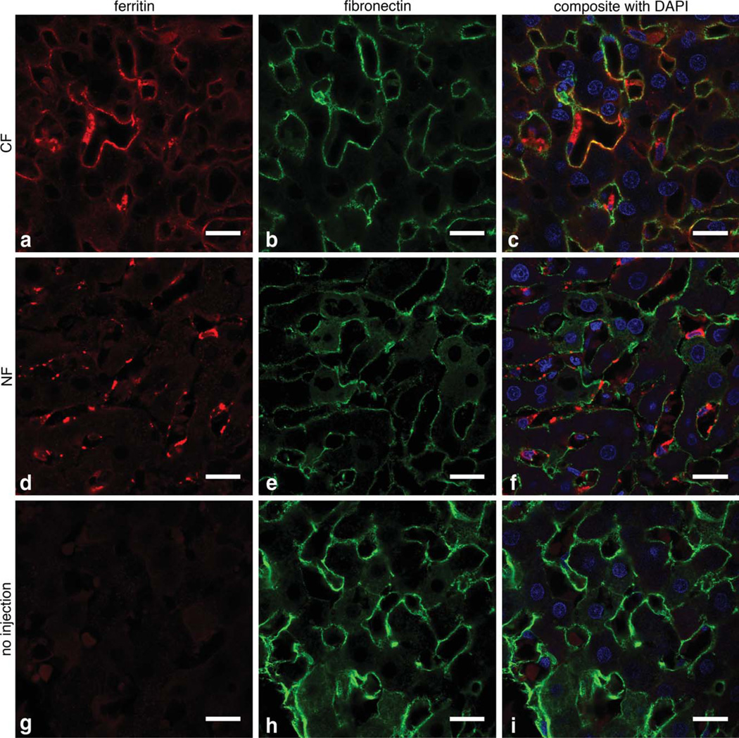
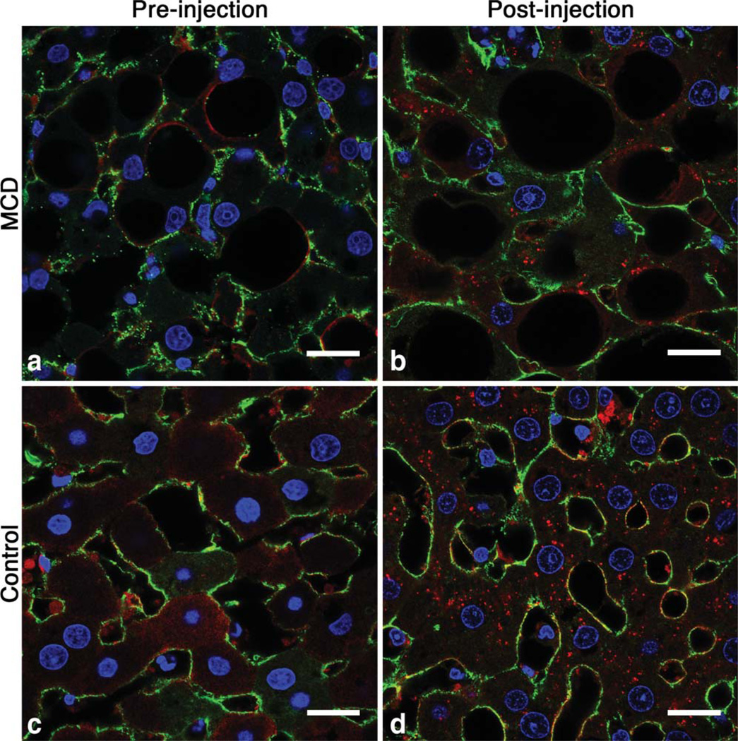
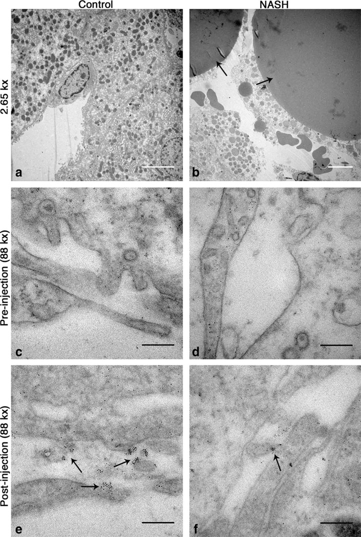
This work demonstrates that the binding of cationized ferritin to the perisinusoidal extracellular matrix is reduced by 55% in a rat model of non-alcoholic steatohepatitis compared to healthy controls. This reduced binding is detectable in vivo with magnetic resonance imaging. Immunofluorescence and electron microscopy indicated that the reduced binding is due to inhibited macromolecular access to the perisinusoidal space caused by non-alcoholic steatohepatitis-related microstructural changes.
The reduced accumulation of intravenously injected cationized ferritin may report on changes in macromolecular access to the liver parenchyma in chronic liver disease.
本研究旨在利用磁共振成像检测肝脏疾病相关的微观结构变化。慢性肝病可导致肝脏微观结构改变,减少血浆进入肝血窦周隙——血浆与肝实质进行交换的部位。血浆进入肝血窦周隙减少会抑制肝功能,导致每年约30000例与慢性肝病相关的死亡。
将细胞外基质特异性阳离子铁蛋白磁共振成像探针静脉注射到健康大鼠和慢性肝病非酒精性脂肪性肝炎大鼠模型体内。随后用T2*加权磁共振成像对大鼠进行成像。
本研究表明,与健康对照组相比,在非酒精性脂肪性肝炎大鼠模型中,阳离子铁蛋白与肝血窦周隙细胞外基质的结合减少了55%。这种结合减少可通过磁共振成像在体内检测到。免疫荧光和电子显微镜检查表明,结合减少是由于非酒精性脂肪性肝炎相关的微观结构变化导致大分子进入肝血窦周隙受到抑制。
静脉注射阳离子铁蛋白的蓄积减少可能反映了慢性肝病中大分子进入肝实质的变化。