Advanced Cardiovascular Imaging Laboratory, Cardiovascular and Pulmonary Branch, Department of Health and Human Services, National Heart, Lung and Blood Institute, National Institutes of Health, Bethesda, MD, USA.
Int J Cardiovasc Imaging. 2013 Jun;29(5):1167-75. doi: 10.1007/s10554-013-0190-1. Epub 2013 Feb 13.
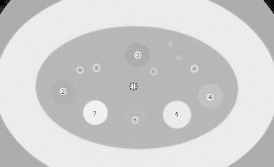
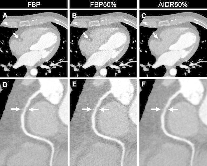
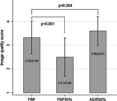
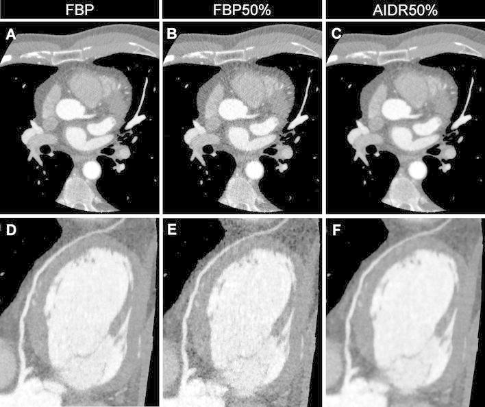
To compare the image quality of coronary CT angiography (CTA) studies between standard filtered back projection (FBP) and adaptive iterative dose reduction in three-dimensions (AIDR3D) reconstruction using CT noise additional software to simulate reduced radiation exposure. Images from 93 consecutive clinical coronary CTA studies were processed utilizing standard FBP, FBP with 50% simulated dose reduction (FBP50%), and AIDR3D with simulated 50% dose reduction (AIDR50%). Signal-to-noise ratio (SNR) and contrast-to-noise ratio (CNR) were measured within 5 regions-of-interest, and image quality for each reconstruction strategy was assessed by two independent readers using a 4-point scale. Compared to FBP, the SNR measured from the AIDR50% images was similar or higher (airway: 38.3 ± 12.7 vs. 38.5 ± 14.5, p = 0.81, fat: 5.5 ± 1.9 vs. 5.4 ± 2.0, p = 0.20, muscle: 3.2 ± 1.2 vs. 3.1 ± 1.3, p = 0.38, aorta: 22.6 ± 9.4 vs. 20.2 ± 9.7, p < 0.0001, liver: 2.7 ± 1.0 vs. 2.3 ± 1.1, p < 0.0001), while the SNR of the FBP50 % images were all lower (p values < 0.0001). The CNR measured from AIDR50% images was also higher than that from the FBP images for the aorta relative to muscle (20.5 ± 9.0 vs. 18.3 ± 9.2, p < 0.0001). The interobserver agreement in the image quality score was excellent (κ = 0.82). The quality score was significantly higher for the AIDR50% images compared to the FBP images (3.6 ± 0.6 vs. 3.3 ± 0.7, p = 0.004). Simulated radiation dose reduction applied to clinical coronary CTA images suggests that a 50% reduction in radiation dose can be achieved with adaptive iterative dose reduction software with image quality that is at least comparable to images acquired at standard radiation exposure and reconstructed with filtered back projection.
为了比较标准滤波反投影(FBP)和三维自适应迭代剂量降低(AIDR3D)重建技术在模拟降低辐射剂量的 CT 噪声附加软件下冠状动脉 CT 血管造影(CTA)研究的图像质量,我们对 93 例连续的临床冠状动脉 CTA 研究的图像进行了处理,分别采用标准 FBP、50%模拟剂量降低的 FBP(FBP50%)和 50%模拟剂量降低的 AIDR3D(AIDR50%)进行处理。在 5 个感兴趣区测量信噪比(SNR)和对比噪声比(CNR),并由两名独立的观察者使用 4 分制评估每种重建策略的图像质量。与 FBP 相比,AIDR50%图像的 SNR 相似或更高(气道:38.3±12.7 与 38.5±14.5,p=0.81,脂肪:5.5±1.9 与 5.4±2.0,p=0.20,肌肉:3.2±1.2 与 3.1±1.3,p=0.38,主动脉:22.6±9.4 与 20.2±9.7,p<0.0001,肝脏:2.7±1.0 与 2.3±1.1,p<0.0001),而 FBP50%图像的 SNR 均较低(p 值<0.0001)。与 FBP 图像相比,AIDR50%图像的主动脉相对于肌肉的 CNR 也更高(20.5±9.0 与 18.3±9.2,p<0.0001)。观察者间图像质量评分的一致性极好(κ=0.82)。与 FBP 图像相比,AIDR50%图像的质量评分显著更高(3.6±0.6 与 3.3±0.7,p=0.004)。应用于临床冠状动脉 CTA 图像的模拟辐射剂量降低表明,使用自适应迭代剂量降低软件可实现 50%的辐射剂量降低,且图像质量至少与标准辐射暴露采集并采用滤波反投影重建的图像相当。