Feger Sarah, Rief Matthias, Zimmermann Elke, Martus Peter, Schuijf Joanne Désirée, Blobel Jörg, Richter Felicitas, Dewey Marc
Department of Radiology, Charité Medical School, Humboldt-Universität zu Berlin, Freie Universität Berlin, Berlin, Germany.
Department of Radiology, Charité Medical School, Berlin, Germany.
PLoS One. 2015 May 6;10(5):e0125943. doi: 10.1371/journal.pone.0125943. eCollection 2015.
The aim of this study was the systematic image quality evaluation of coronary CT angiography (CTA), reconstructed with the 3 different levels of adaptive iterative dose reduction (AIDR 3D) and compared to filtered back projection (FBP) with quantum denoising software (QDS).
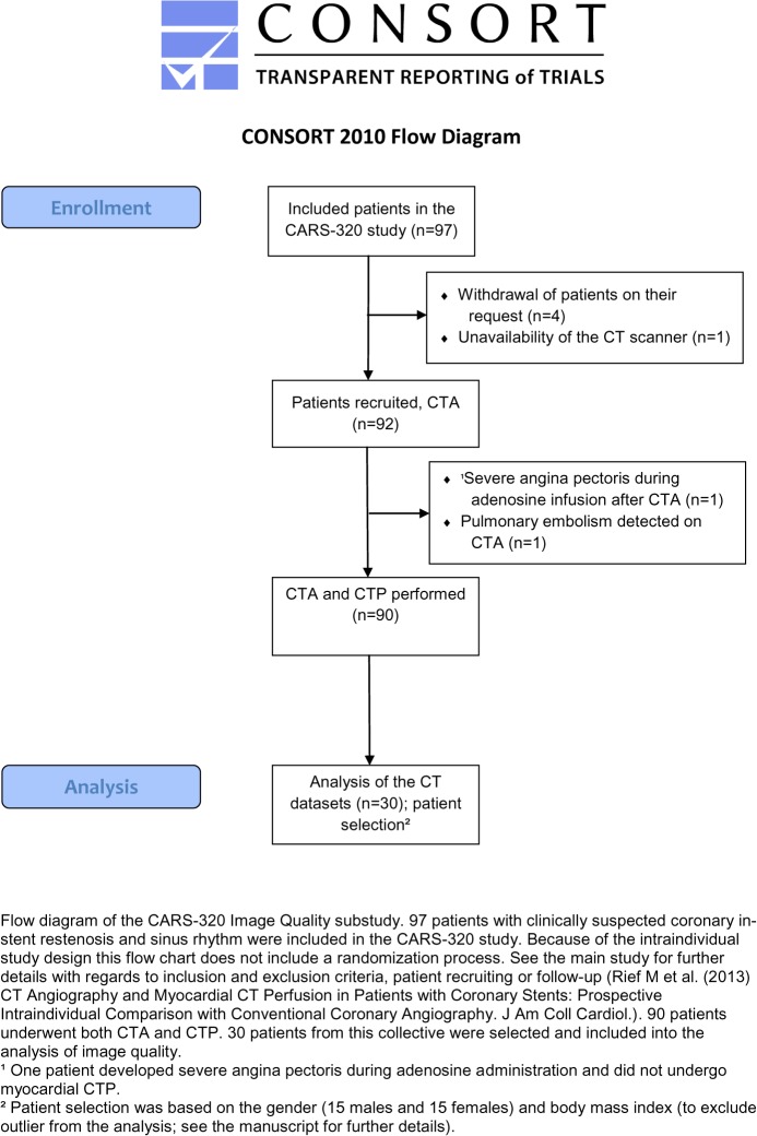
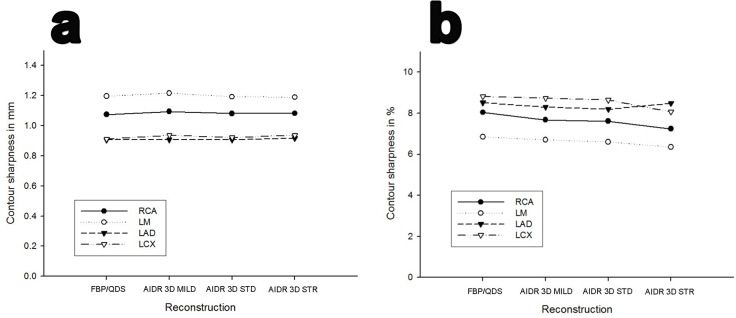
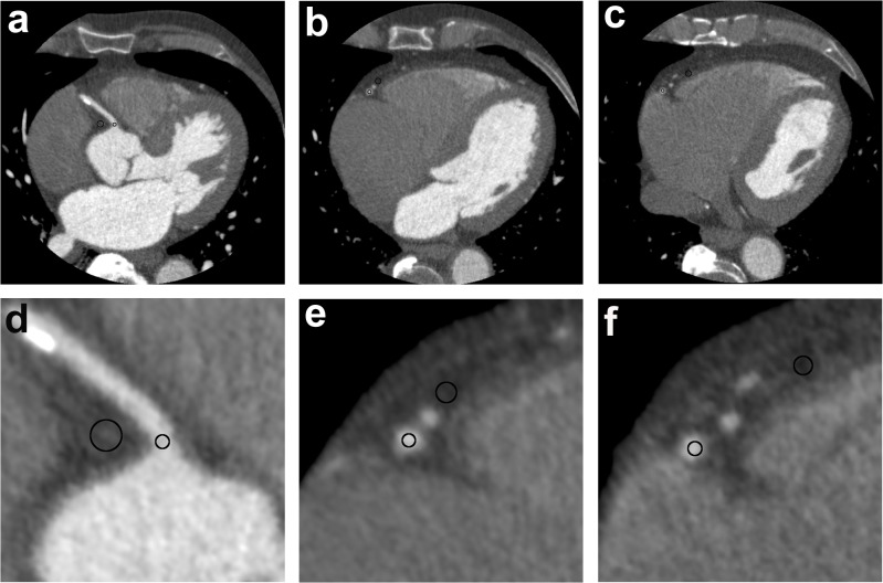
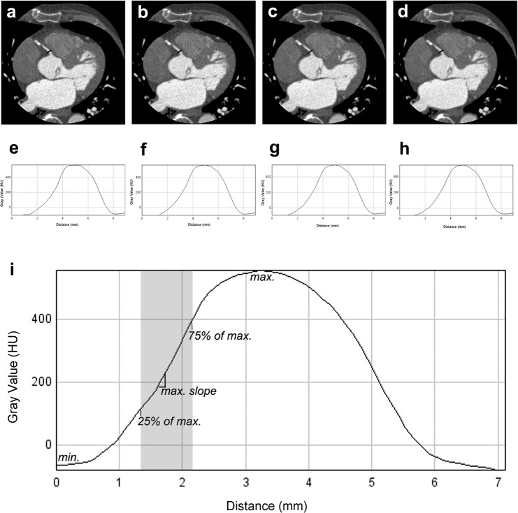
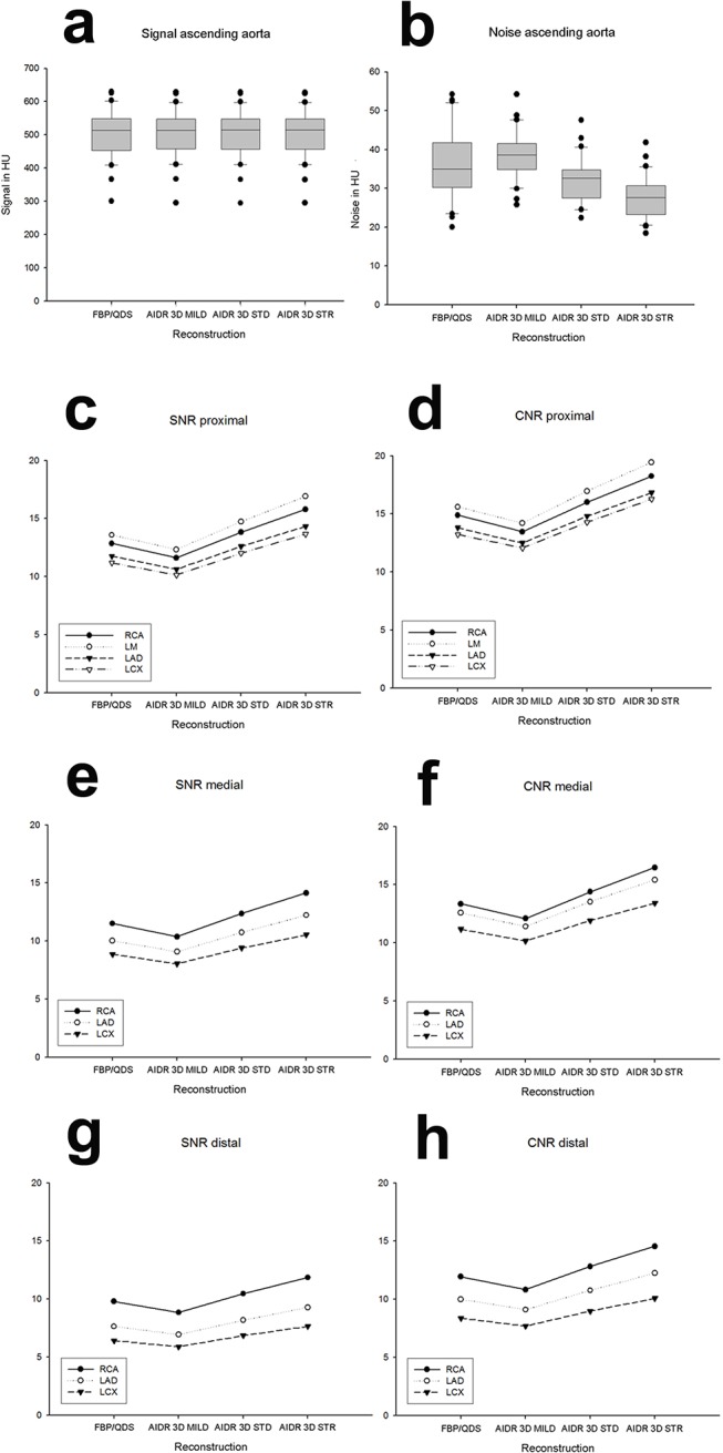
Standard-dose CTA raw data of 30 patients with mean radiation dose of 3.2 ± 2.6 mSv were reconstructed using AIDR 3D mild, standard, strong and compared to FBP/QDS. Objective image quality comparison (signal, noise, signal-to-noise ratio (SNR), contrast-to-noise ratio (CNR), contour sharpness) was performed using 21 measurement points per patient, including measurements in each coronary artery from proximal to distal.
Objective image quality parameters improved with increasing levels of AIDR 3D. Noise was lowest in AIDR 3D strong (p ≤ 0.001 at 20/21 measurement points; compared with FBP/QDS). Signal and contour sharpness analysis showed no significant difference between the reconstruction algorithms for most measurement points. Best coronary SNR and CNR were achieved with AIDR 3D strong. No loss of SNR or CNR in distal segments was seen with AIDR 3D as compared to FBP.
On standard-dose coronary CTA images, AIDR 3D strong showed higher objective image quality than FBP/QDS without reducing contour sharpness.
Clinicaltrials.gov NCT00967876.
本研究旨在对采用3种不同级别自适应迭代剂量降低(AIDR 3D)重建的冠状动脉CT血管造影(CTA)进行系统的图像质量评估,并与使用量子降噪软件(QDS)的滤波反投影(FBP)进行比较。
对30例平均辐射剂量为3.2±2.6 mSv的患者的标准剂量CTA原始数据采用轻度、标准、重度AIDR 3D进行重建,并与FBP/QDS进行比较。使用每位患者21个测量点进行客观图像质量比较(信号、噪声、信噪比(SNR)、对比噪声比(CNR)、轮廓清晰度),包括从近端到远端对每条冠状动脉进行测量。
随着AIDR 3D级别增加,客观图像质量参数得到改善。重度AIDR 3D的噪声最低(21个测量点中的20个点p≤0.001;与FBP/QDS相比)。信号和轮廓清晰度分析显示,大多数测量点的重建算法之间无显著差异。重度AIDR 3D实现了最佳的冠状动脉SNR和CNR。与FBP相比,AIDR 3D在远端节段未见SNR或CNR损失。
在标准剂量冠状动脉CTA图像上,重度AIDR 3D显示出比FBP/QDS更高的客观图像质量,且未降低轮廓清晰度。
Clinicaltrials.gov NCT00967876。