• 文献检索
  • 文档翻译
  • 深度研究
  • 学术资讯
  • Suppr Zotero 插件Zotero 插件
  • 邀请有礼
  • 套餐&价格
  • 历史记录
应用&插件
Suppr Zotero 插件Zotero 插件浏览器插件Mac 客户端Windows 客户端微信小程序
定价
高级版会员购买积分包购买API积分包
服务
文献检索文档翻译深度研究API 文档MCP 服务
关于我们
关于 Suppr公司介绍联系我们用户协议隐私条款
关注我们

Suppr 超能文献

核心技术专利:CN118964589B侵权必究
粤ICP备2023148730 号-1Suppr @ 2026

文献检索

告别复杂PubMed语法,用中文像聊天一样搜索,搜遍4000万医学文献。AI智能推荐,让科研检索更轻松。

立即免费搜索

文件翻译

保留排版,准确专业,支持PDF/Word/PPT等文件格式,支持 12+语言互译。

免费翻译文档

深度研究

AI帮你快速写综述,25分钟生成高质量综述,智能提取关键信息,辅助科研写作。

立即免费体验

青光眼检测的三维谱域光学相干断层扫描数据分析。

Three-dimensional spectral-domain optical coherence tomography data analysis for glaucoma detection.

机构信息

University of Pittsburgh Medical Center Eye Center, Eye and Ear Institute, Ophthalmology and Visual Science Research Center, Department of Ophthalmology, University of Pittsburgh School of Medicine, Pittsburgh, Pennsylvania, USA.

出版信息

PLoS One. 2013;8(2):e55476. doi: 10.1371/journal.pone.0055476. Epub 2013 Feb 11.

DOI:10.1371/journal.pone.0055476
PMID:23408988
原文链接:https://pmc.ncbi.nlm.nih.gov/articles/PMC3569462/
Abstract

PURPOSE

To develop a new three-dimensional (3D) spectral-domain optical coherence tomography (SD-OCT) data analysis method using a machine learning technique based on variable-size super pixel segmentation that efficiently utilizes full 3D dataset to improve the discrimination between early glaucomatous and healthy eyes.

METHODS

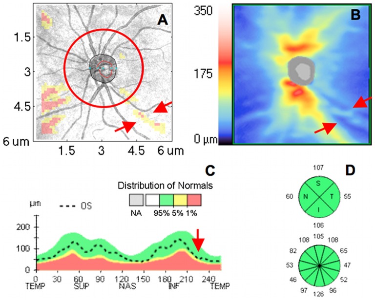
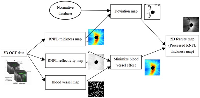
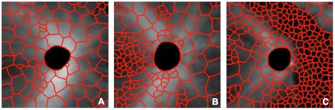
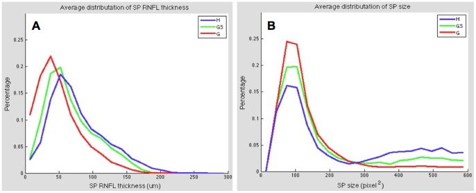
192 eyes of 96 subjects (44 healthy, 59 glaucoma suspect and 89 glaucomatous eyes) were scanned with SD-OCT. Each SD-OCT cube dataset was first converted into 2D feature map based on retinal nerve fiber layer (RNFL) segmentation and then divided into various number of super pixels. Unlike the conventional super pixel having a fixed number of points, this newly developed variable-size super pixel is defined as a cluster of homogeneous adjacent pixels with variable size, shape and number. Features of super pixel map were extracted and used as inputs to machine classifier (LogitBoost adaptive boosting) to automatically identify diseased eyes. For discriminating performance assessment, area under the curve (AUC) of the receiver operating characteristics of the machine classifier outputs were compared with the conventional circumpapillary RNFL (cpRNFL) thickness measurements.

RESULTS

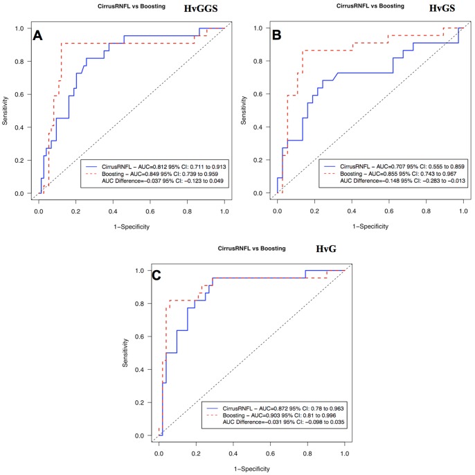
The super pixel analysis showed statistically significantly higher AUC than the cpRNFL (0.855 vs. 0.707, respectively, p = 0.031, Jackknife test) when glaucoma suspects were discriminated from healthy, while no significant difference was found when confirmed glaucoma eyes were discriminated from healthy eyes.

CONCLUSIONS

A novel 3D OCT analysis technique performed at least as well as the cpRNFL in glaucoma discrimination and even better at glaucoma suspect discrimination. This new method has the potential to improve early detection of glaucomatous damage.

摘要

目的

开发一种新的基于机器的三维(3D)光谱域光相干断层扫描(SD-OCT)数据分析方法,该方法基于可变大小超像素分割的机器学习技术,有效地利用全 3D 数据集,提高早期青光眼和正常眼之间的区分能力。

方法

对 96 名患者(44 名正常、59 名青光眼疑似和 89 名青光眼患者)的 192 只眼进行了 SD-OCT 扫描。每个 SD-OCT 立方数据集首先基于视网膜神经纤维层(RNFL)分割转换为 2D 特征图,然后分为不同数量的超像素。与传统的具有固定点数的超像素不同,新开发的可变大小超像素定义为具有可变大小、形状和数量的同质相邻像素的簇。提取超像素图的特征,并将其作为输入提供给机器分类器(LogitBoost 自适应增强),以自动识别患病眼。为了进行判别性能评估,比较了机器分类器输出的接收者操作特性曲线下的面积(AUC)与传统的周边视网膜神经纤维层(cpRNFL)厚度测量值。

结果

超像素分析显示,在区分青光眼疑似患者和正常患者时,其 AUC 显著高于 cpRNFL(分别为 0.855 和 0.707,p=0.031,Jackknife 检验),而在区分确诊青光眼患者和正常患者时,两者之间无显著差异。

结论

一种新的 3D OCT 分析技术在青光眼鉴别中的表现至少与 cpRNFL 一样好,甚至在青光眼疑似患者鉴别中表现更好。这种新方法有可能提高青光眼损伤的早期检测能力。

https://cdn.ncbi.nlm.nih.gov/pmc/blobs/728e/3569462/2b2f656c1717/pone.0055476.g006.jpg
https://cdn.ncbi.nlm.nih.gov/pmc/blobs/728e/3569462/4a54213c3ced/pone.0055476.g001.jpg
https://cdn.ncbi.nlm.nih.gov/pmc/blobs/728e/3569462/c73e2a22b1b8/pone.0055476.g002.jpg
https://cdn.ncbi.nlm.nih.gov/pmc/blobs/728e/3569462/6d85ec2e4ce7/pone.0055476.g003.jpg
https://cdn.ncbi.nlm.nih.gov/pmc/blobs/728e/3569462/2f02b5b41bcd/pone.0055476.g004.jpg
https://cdn.ncbi.nlm.nih.gov/pmc/blobs/728e/3569462/570c91bdac73/pone.0055476.g005.jpg
https://cdn.ncbi.nlm.nih.gov/pmc/blobs/728e/3569462/2b2f656c1717/pone.0055476.g006.jpg

相似文献

1
Three-dimensional spectral-domain optical coherence tomography data analysis for glaucoma detection.青光眼检测的三维谱域光学相干断层扫描数据分析。
PLoS One. 2013;8(2):e55476. doi: 10.1371/journal.pone.0055476. Epub 2013 Feb 11.
2
3D optical coherence tomography super pixel with machine classifier analysis for glaucoma detection.用于青光眼检测的基于机器分类器分析的三维光学相干断层扫描超像素技术
Annu Int Conf IEEE Eng Med Biol Soc. 2011;2011:3395-8. doi: 10.1109/IEMBS.2011.6090919.
3
Diagnostic ability of macular ganglion cell inner plexiform layer measurements in glaucoma using swept source and spectral domain optical coherence tomography.使用扫频源和谱域光学相干断层扫描测量黄斑神经节细胞内丛状层在青光眼诊断中的能力
PLoS One. 2015 May 15;10(5):e0125957. doi: 10.1371/journal.pone.0125957. eCollection 2015.
4
The ability of macular parameters and circumpapillary retinal nerve fiber layer by three SD-OCT instruments to diagnose highly myopic glaucoma.三种 SD-OCT 仪器的黄斑参数和环周视网膜神经纤维层对高度近视性青光眼的诊断能力。
Invest Ophthalmol Vis Sci. 2013 Sep 5;54(9):6025-32. doi: 10.1167/iovs.13-12630.
5
Retinal Nerve Fiber Layer Features Identified by Unsupervised Machine Learning on Optical Coherence Tomography Scans Predict Glaucoma Progression.基于光学相干断层扫描的无监督机器学习识别的视网膜神经纤维层特征可预测青光眼进展。
Invest Ophthalmol Vis Sci. 2018 Jun 1;59(7):2748-2756. doi: 10.1167/iovs.17-23387.
6
Assessment of a Segmentation-Free Deep Learning Algorithm for Diagnosing Glaucoma From Optical Coherence Tomography Scans.基于无分割深度学习算法的光学相干断层扫描图像青光眼诊断评估。
JAMA Ophthalmol. 2020 Apr 1;138(4):333-339. doi: 10.1001/jamaophthalmol.2019.5983.
7
Comparative assessment for the ability of Cirrus, RTVue, and 3D-OCT to diagnose glaucoma.比较评估 Cirrus、RTVue 和 3D-OCT 诊断青光眼的能力。
Invest Ophthalmol Vis Sci. 2013 Jul 10;54(7):4478-84. doi: 10.1167/iovs.12-11268.
8
Retinal Nerve Fiber Layer Thickness Measurement Comparison Using Spectral Domain and Swept Source Optical Coherence Tomography.使用光谱域和扫频光学相干断层扫描技术比较视网膜神经纤维层厚度测量
Korean J Ophthalmol. 2016 Apr;30(2):140-7. doi: 10.3341/kjo.2016.30.2.140. Epub 2016 Mar 25.
9
Diagnosing Glaucoma With Spectral-Domain Optical Coherence Tomography Using Deep Learning Classifier.使用深度学习分类器的谱域光相干断层扫描诊断青光眼。
J Glaucoma. 2020 Apr;29(4):287-294. doi: 10.1097/IJG.0000000000001458.
10
Glaucoma discrimination of segmented cirrus spectral domain optical coherence tomography (SD-OCT) macular scans.节段性脉络膜 SD-OCT 黄斑扫描的青光眼鉴别。
Br J Ophthalmol. 2012 Nov;96(11):1420-5. doi: 10.1136/bjophthalmol-2011-301021. Epub 2012 Aug 22.

引用本文的文献

1
OCT-based diagnosis of glaucoma and glaucoma stages using explainable machine learning.基于光学相干断层扫描(OCT),利用可解释机器学习进行青光眼及青光眼分期的诊断。
Sci Rep. 2025 Jan 28;15(1):3592. doi: 10.1038/s41598-025-87219-w.
2
Artificial intelligence applications in ophthalmic optical coherence tomography: a 12-year bibliometric analysis.人工智能在眼科光学相干断层扫描中的应用:一项为期12年的文献计量分析。
Int J Ophthalmol. 2024 Dec 18;17(12):2295-2307. doi: 10.18240/ijo.2024.12.19. eCollection 2024.
3
The utilization of artificial intelligence in glaucoma: diagnosis versus screening.

本文引用的文献

1
Optical coherence tomography: future trends for imaging in glaucoma.光学相干断层扫描:青光眼成像的未来趋势
Optom Vis Sci. 2012 May;89(5):E554-62. doi: 10.1097/OPX.0b013e31824eeb43.
2
Optical coherence tomography: history, current status, and laboratory work.光学相干断层扫描:历史、现状和实验室工作。
Invest Ophthalmol Vis Sci. 2011 Apr 14;52(5):2425-36. doi: 10.1167/iovs.10-6312. Print 2011 Apr.
3
Retinal nerve fiber layer imaging with spectral-domain optical coherence tomography: analysis of the retinal nerve fiber layer map for glaucoma detection.
人工智能在青光眼领域的应用:诊断与筛查
Front Ophthalmol (Lausanne). 2024 Mar 6;4:1368081. doi: 10.3389/fopht.2024.1368081. eCollection 2024.
4
Diagnosing glaucoma in primary eye care and the role of Artificial Intelligence applications for reducing the prevalence of undetected glaucoma in Australia.初级眼保健中的青光眼诊断和人工智能应用在降低澳大利亚未检出青光眼患病率中的作用。
Eye (Lond). 2024 Aug;38(11):2003-2013. doi: 10.1038/s41433-024-03026-z. Epub 2024 Mar 21.
5
On Machine Learning in Clinical Interpretation of Retinal Diseases Using OCT Images.关于使用光学相干断层扫描(OCT)图像进行视网膜疾病临床解读的机器学习
Bioengineering (Basel). 2023 Mar 24;10(4):407. doi: 10.3390/bioengineering10040407.
6
Clinical Applications of Artificial Intelligence in Glaucoma.人工智能在青光眼领域的临床应用
J Ophthalmic Vis Res. 2023 Feb 21;18(1):97-112. doi: 10.18502/jovr.v18i1.12730. eCollection 2023 Jan-Mar.
7
Depth-resolved extraction of optical attenuation for glaucoma assessment in clinical settings: a pilot study.临床环境中用于青光眼评估的光学衰减深度分辨提取:一项初步研究
Biomed Opt Express. 2022 Jul 26;13(8):4326-4337. doi: 10.1364/BOE.461348. eCollection 2022 Aug 1.
8
Artificial Intelligence Algorithms to Diagnose Glaucoma and Detect Glaucoma Progression: Translation to Clinical Practice.用于诊断青光眼和检测青光眼病情进展的人工智能算法:向临床实践的转化
Transl Vis Sci Technol. 2020 Oct 15;9(2):55. doi: 10.1167/tvst.9.2.55. eCollection 2020 Oct.
9
Current applications of machine learning in the screening and diagnosis of glaucoma: a systematic review and Meta-analysis.机器学习在青光眼筛查与诊断中的当前应用:系统评价与Meta分析
Int J Ophthalmol. 2020 Jan 18;13(1):149-162. doi: 10.18240/ijo.2020.01.22. eCollection 2020.
10
Joint retina segmentation and classification for early glaucoma diagnosis.用于早期青光眼诊断的联合视网膜分割与分类
Biomed Opt Express. 2019 Apr 30;10(5):2639-2656. doi: 10.1364/BOE.10.002639. eCollection 2019 May 1.
频域光学相干断层扫描的视网膜神经纤维层成像:用于青光眼检测的视网膜神经纤维层图分析。
Ophthalmology. 2010 Sep;117(9):1684-91. doi: 10.1016/j.ophtha.2010.01.026. Epub 2010 Jul 21.
4
Three dimensional optical coherence tomography imaging: advantages and advances.三维光学相干断层成像:优势与进展。
Prog Retin Eye Res. 2010 Nov;29(6):556-79. doi: 10.1016/j.preteyeres.2010.05.005. Epub 2010 Jun 11.
5
Spectral domain optical coherence tomography for glaucoma (an AOS thesis).用于青光眼的光谱域光学相干断层扫描(一篇应用光学与光子学理学硕士论文)
Trans Am Ophthalmol Soc. 2008;106:426-58.
6
Imaging of the retinal nerve fibre layer for glaucoma.青光眼视网膜神经纤维层成像
Br J Ophthalmol. 2009 Feb;93(2):139-43. doi: 10.1136/bjo.2008.145540. Epub 2008 Nov 21.
7
Automated optic disk boundary detection by modified active contour model.基于改进活动轮廓模型的自动视盘边界检测
IEEE Trans Biomed Eng. 2007 Mar;54(3):473-82. doi: 10.1109/TBME.2006.888831.
8
Relationship between visual field sensitivity and retinal nerve fiber layer thickness as measured by optical coherence tomography.通过光学相干断层扫描测量的视野敏感度与视网膜神经纤维层厚度之间的关系。
Invest Ophthalmol Vis Sci. 2007 Jan;48(1):258-63. doi: 10.1167/iovs.06-0410.
9
Macular segmentation with optical coherence tomography.利用光学相干断层扫描进行黄斑分割
Invest Ophthalmol Vis Sci. 2005 Jun;46(6):2012-7. doi: 10.1167/iovs.04-0335.
10
Ultrahigh-resolution ophthalmic optical coherence tomography.超高分辨率眼科光学相干断层扫描
Nat Med. 2001 Apr;7(4):502-7. doi: 10.1038/86589.