Department of Pathology, Faculty of Veterinary Science, Chulalongkorn University, Henri-Dunant Rd, Bangkok 10330, Thailand.
Virol J. 2013 Mar 16;10:88. doi: 10.1186/1743-422X-10-88.
Following the emergence of the pandemic H1N1 influenza A virus in 2009 in humans, this novel virus spread into the swine population. Pigs represent a potential host for this virus and can serve as a mixing vessel for genetic mutations of the influenza virus. Reassortant viruses eventually emerged from the 2009 pandemic and were reported in swine populations worldwide including Thailand. As a result of the discovery of this emergent disease, pathogenesis studies of this novel virus were conducted in order that future disease protection and control measures in swine and human populations could be enacted.
The pandemic H1N1 2009 virus (pH1N1) and its reassortant virus (rH1N1) isolated from pigs in Thailand were inoculated into 2 separate cohorts of 9, 3-week-old pigs. Cohorts were consisted of one group experimentally infected with pH1N1 and one group with rH1N1. A negative control group consisting of 3 pigs was also included. Clinical signs, viral shedding and pathological lesions were investigated and compared. Later, 3 pigs from viral inoculated groups and 1 pig from the control group were necropsied at 2, 4, and 12 days post inoculation (DPI).
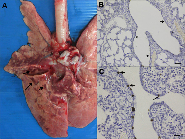
The results indicated that pigs infected with both viruses demonstrated typical flu-like clinical signs and histopathological lesions of varying severity. Influenza infected-pigs of both groups had mild to moderate pulmonary signs on 1-4 DPI. Interestingly, pigs in both groups demonstrated viral RNA detection in the nasal swabs until the end of the experiment (12 DPI).
The present study demonstrated that both the pH1N1 and rH1N1 influenza viruses, isolated from naturally infected pigs, induced acute respiratory disease in experimentally inoculated nursery pigs. Although animals in the rH1N1-infected cohort demonstrated more severe clinical signs, had higher numbers of pigs shedding the virus, were noted to have increased histopathological severity of lung lesions and increased viral antigen in lung tissue, the findings were not statistically significant in comparison with the pH1N1-infected group. Interestingly, viral genetic material of both viruses could be detected from the nasal swabs until the end of the experiment. Similar to other swine influenza viruses, the clinical signs and pathological lesions in both rH1N1 and pH1N1 were limited to the respiratory tract.
2009 年大流行 H1N1 流感病毒在人类中出现后,这种新型病毒传播到猪群中。猪是这种病毒的潜在宿主,并且可以作为流感病毒遗传突变的混合容器。重组病毒最终从 2009 年大流行中出现,并在包括泰国在内的全球猪群中报告。由于发现了这种新出现的疾病,因此对这种新型病毒进行了发病机制研究,以便可以对猪和人群实施未来的疾病预防和控制措施。
从泰国猪中分离出的大流行 H1N1 2009 病毒(pH1N1)和其重组病毒(rH1N1)分别接种到两组 9 只,3 周龄的猪中。两组分别包括一组用 pH1N1 感染的实验组和一组用 rH1N1 感染的实验组。还包括一组由 3 只猪组成的阴性对照组。对临床症状,病毒脱落和病理损伤进行了调查和比较。之后,在接种病毒后的两组中各有 3 只猪和对照组中的 1 只猪在接种后 2、4 和 12 天(DPI)进行剖检。
结果表明,感染两种病毒的猪均表现出典型的流感样临床症状和不同严重程度的组织病理学病变。两组感染流感的猪在 1-4 DPI 时出现轻度至中度肺部症状。有趣的是,两组猪在实验结束时(12 DPI)均在鼻拭子中检测到病毒 RNA。
本研究表明,从自然感染的猪中分离出的 pH1N1 和 rH1N1 流感病毒均在实验接种的仔猪中引起急性呼吸道疾病。尽管 rH1N1 感染组的动物表现出更严重的临床症状,有更多的猪排出病毒,肺部病变的组织病理学严重程度更高,肺部组织中的病毒抗原增加,但与 pH1N1 感染组相比,这些发现没有统计学意义。有趣的是,两种病毒的病毒遗传物质均可从鼻拭子中检测到,直到实验结束。与其他猪流感病毒一样,rH1N1 和 pH1N1 的临床症状和病理损伤仅限于呼吸道。