Department of Comparative Pathology, Veterinary Faculty, University of Córdoba-Agrifood Campus of International Excellence (ceiA3), Edificio Sanidad Animal, Campus de Rabanales, Córdoba 14014, Spain.
Vet Res. 2013 Mar 18;44(1):20. doi: 10.1186/1297-9716-44-20.
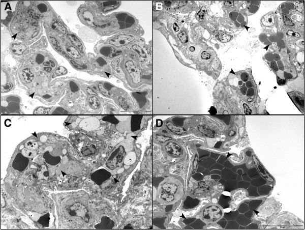
Resistance to respiratory disease in cattle requires host defense mechanisms that protect against pathogens which have evolved sophisticated strategies to evade them, including an altered function of pulmonary macrophages (MΦs) or the induction of inflammatory responses that cause lung injury and sepsis. The aim of this study was to clarify the mechanisms responsible for vascular changes occurring in the lungs of calves infected with bovine viral diarrhea virus (BVDV) and challenged later with bovine herpesvirus type 1 (BHV-1), evaluating the role of MΦs in the development of pathological lesions in this organ. For this purpose, pulmonary lesions were compared between co-infected calves and healthy animals inoculated only with BHV-1 through immunohistochemical (MAC387, TNFα, IL-1α, iNOS, COX-2 and Factor-VIII) and ultrastructural studies. Both groups of calves presented important vascular alterations produced by fibrin microthrombi and platelet aggregations within the blood vessels. These findings were earlier and more severe in the co-infected group, indicating that the concomitance of BVDV and BHV-1 in the lungs disrupts the pulmonary homeostasis by facilitating the establishment of an inflammatory and procoagulant environment modulated by inflammatory mediators released by pulmonary MΦs. In this regard, the co-infected calves, in spite of presenting a greater number of IMΦs than single-infected group, show a significant decrease in iNOS expression coinciding with the presence of more coagulation lesions. Moreover, animals pre-inoculated with BVDV displayed an alteration in the response of pro-inflammatory cytokines (TNFα and IL-1), which play a key role in activating the immune response, as well as in the local cell-mediated response.
牛的呼吸道疾病抗性需要宿主防御机制来保护其免受已进化出复杂策略来逃避这些防御机制的病原体的侵害,包括肺巨噬细胞(MΦ)功能改变或引发肺损伤和败血症的炎症反应的诱导。本研究旨在阐明牛病毒性腹泻病毒(BVDV)感染牛犊后肺部发生血管变化的机制,并随后用牛疱疹病毒 1 型(BHV-1)对其进行挑战,评估 MΦ 在该器官病理损伤发展中的作用。为此,通过免疫组织化学(MAC387、TNFα、IL-1α、iNOS、COX-2 和因子 VIII)和超微结构研究比较了同时感染的牛犊和仅接种 BHV-1 的健康动物的肺部病变。两组牛犊的肺部均出现重要的血管改变,由血管内的纤维蛋白微血栓和血小板聚集引起。在同时感染组中,这些发现更早且更严重,表明 BVDV 和 BHV-1 在肺部的共存通过促进由肺 MΦ 释放的炎症介质调节的炎症和促凝环境的建立来破坏肺内稳态。在这方面,尽管同时感染的牛犊比单一感染组有更多的 IMΦ,但 iNOS 表达显著下降,同时伴有更多的凝血病变。此外,预先接种 BVDV 的动物表现出促炎细胞因子(TNFα 和 IL-1)反应的改变,这些细胞因子在激活免疫反应以及局部细胞介导反应中起着关键作用。