Laboratory of Cutaneous Physiopathology, San Gallicano Dermatologic Institute, Istituto Di Ricovero e Cura a Carattere Scientifico, Rome, Italy.
PLoS One. 2013;8(3):e59782. doi: 10.1371/journal.pone.0059782. Epub 2013 Mar 26.
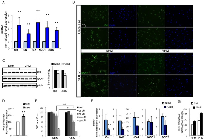
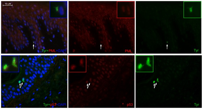
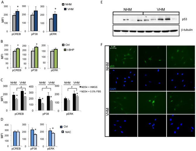
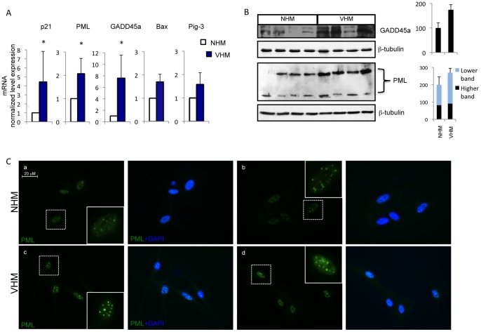
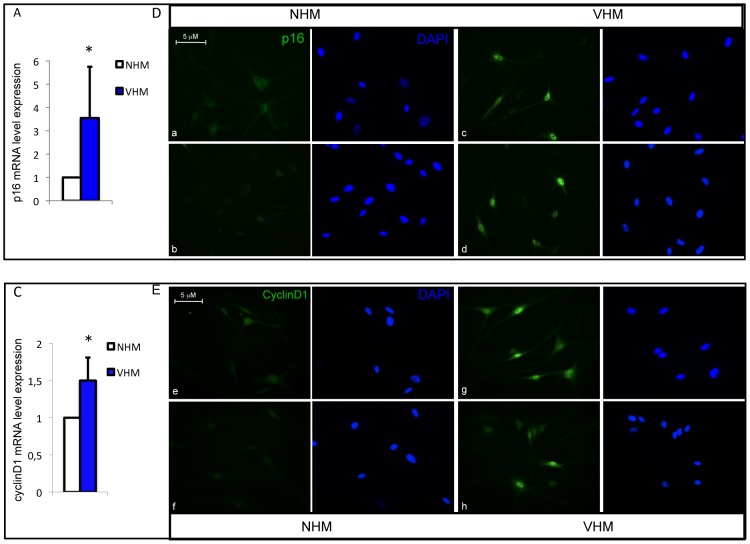
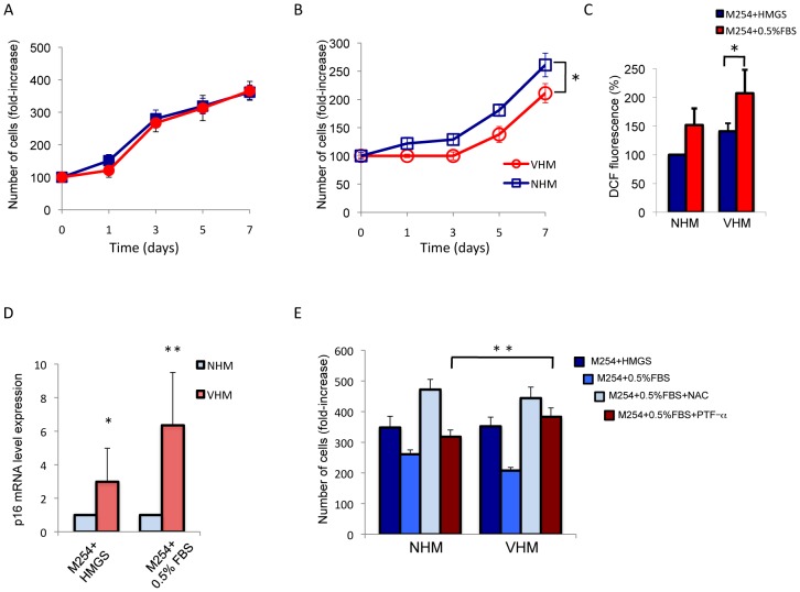
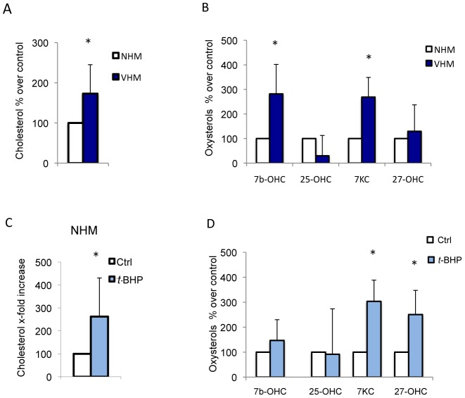
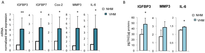
Vitiligo is characterized by the progressive disappearance of pigment cells from skin and hair follicle. Several in vitro and in vivo studies show evidence of an altered redox status, suggesting that loss of cellular redox equilibrium might be the pathogenic mechanism in vitiligo. However, despite the numerous data supporting a pathogenic role of oxidative stress, there is still no consensus explanation underlying the oxidative stress-driven disappear of melanocytes from the epidermis. In this study, in vitro characterization of melanocytes cultures from non-lesional vitiligo skin revealed at the cellular level aberrant function of signal transduction pathways common with neurodegenerative diseases including modification of lipid metabolism, hyperactivation of mitogen-activated protein kinase (MAPK) and cAMP response element-binding protein (CREB), constitutive p53-dependent stress signal transduction cascades, and enhanced sensibility to pro-apoptotic stimuli. Notably, these long-term effects of subcytotoxic oxidative stress are also biomarkers of pre-senescent cellular phenotype. Consistent with this, vitiligo cells showed a significant increase in p16 that did not correlate with the chronological age of the donor. Moreover, vitiligo melanocytes produced many biologically active proteins among the senescence-associated secretory phenotype (SAPS), such as interleukin-6 (IL-6), matrix metallo proteinase-3 (MMP3), cyclooxygenase-2 (Cox-2), insulin-like growth factor-binding protein-3 and 7 (IGFBP3, IGFBP7). Together, these data argue for a complicated pathophysiologic puzzle underlying melanocytes degeneration resembling, from the biological point of view, neurodegenerative diseases. Our results suggest new possible targets for intervention that in combination with current therapies could correct melanocytes intrinsic defects.
白癜风的特征是皮肤和毛囊中的色素细胞逐渐消失。多项体外和体内研究表明存在氧化还原状态改变的证据,这表明细胞氧化还原平衡的丧失可能是白癜风的发病机制。然而,尽管有大量数据支持氧化应激的致病作用,但对于氧化应激驱动黑色素细胞从表皮消失的机制仍没有共识解释。在这项研究中,对非病变性白癜风皮肤来源的黑素细胞培养物进行了体外特征分析,结果表明信号转导通路的异常功能与神经退行性疾病(包括脂质代谢改变、丝裂原活化蛋白激酶(MAPK)和 cAMP 反应元件结合蛋白(CREB)的过度激活、p53 依赖性应激信号转导级联的组成性激活以及对促凋亡刺激的敏感性增强)。值得注意的是,这种亚细胞毒性氧化应激的长期作用也是衰老前细胞表型的生物标志物。与此一致,白癜风细胞中 p16 的表达显著增加,而与供体的实际年龄无关。此外,白癜风黑素细胞产生了许多衰老相关分泌表型(SAPS)中的生物活性蛋白,如白细胞介素 6(IL-6)、基质金属蛋白酶 3(MMP3)、环氧化酶 2(Cox-2)、胰岛素样生长因子结合蛋白 3 和 7(IGFBP3、IGFBP7)。综上所述,这些数据表明黑色素细胞退化的背后存在复杂的病理生理难题,从生物学角度来看,与神经退行性疾病相似。我们的研究结果为干预提供了新的可能靶点,与目前的治疗方法相结合,可以纠正黑色素细胞的内在缺陷。