Department of Pediatrics, Seoul National University Bundang Hospital, Seongnam, South Korea.
Pediatr Res. 2013 Jul;74(1):11-8. doi: 10.1038/pr.2013.58. Epub 2013 Apr 5.
We previously showed that intra-amniotic lipopolysaccharide (LPS) amplifies alveolar hypoplasia induced by postnatal hyperoxia. We determined whether the priming effect of intra-amniotic LPS amplifies hyperoxia-induced airway hyperreactivity (AHR).
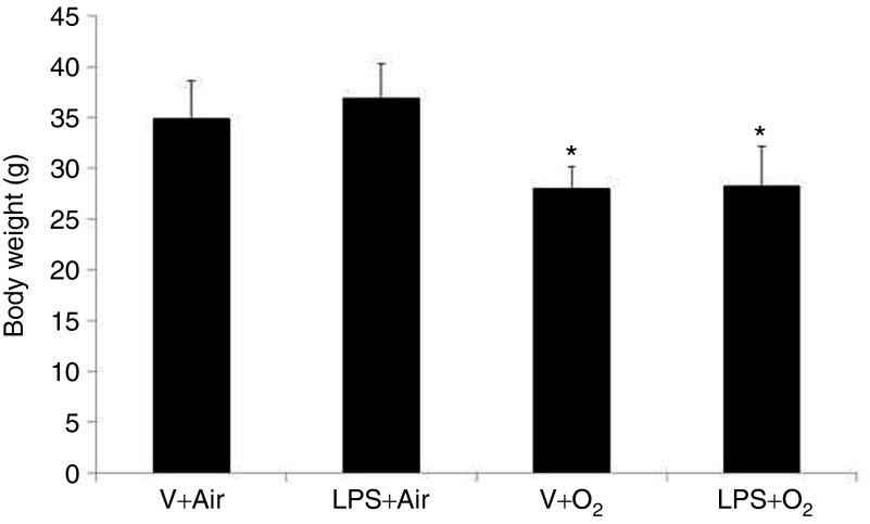
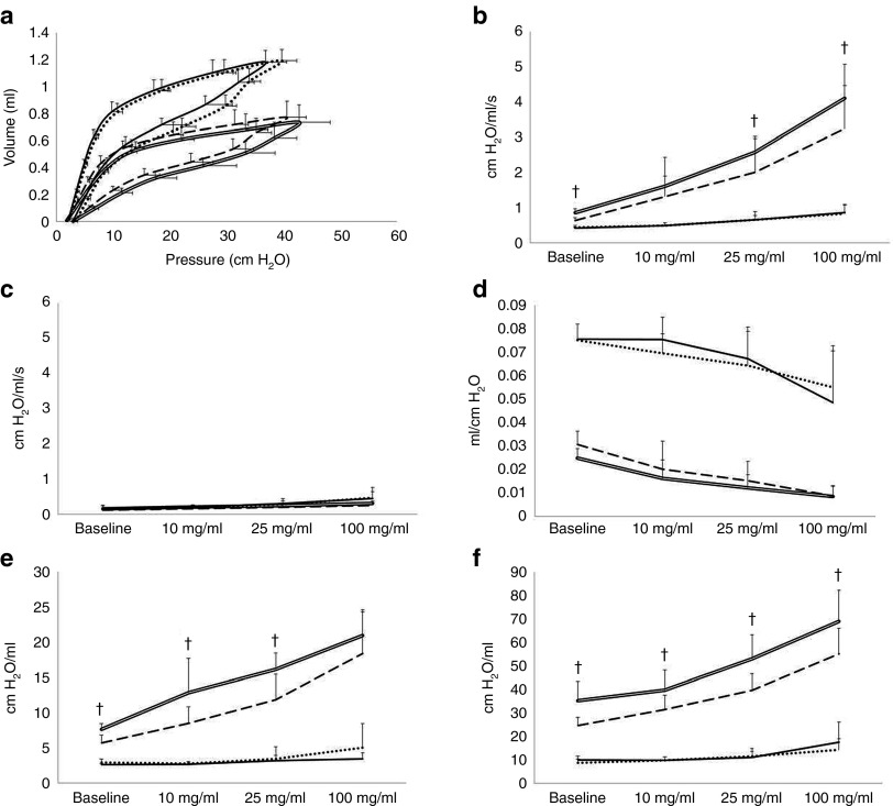
LPS or normal saline was injected into the amniotic cavities of pregnant rats at the 20th day of gestation. After birth, rat pups were exposed to 60% O₂ or air for 14 d. On postnatal day 14, rat pups underwent forced oscillometry, which included a challenge with nebulized methacholine, and the lungs were harvested for morphological studies.
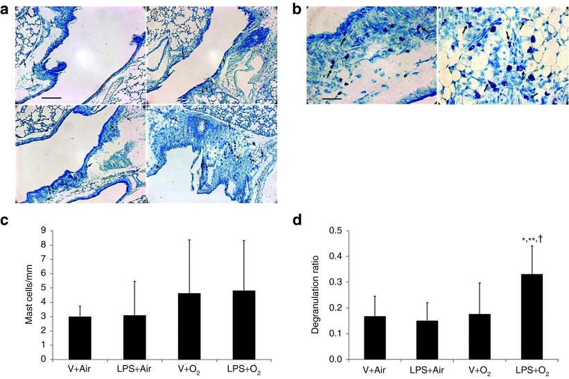
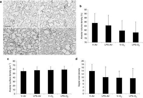
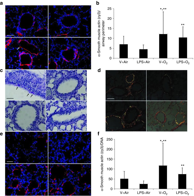
Hyperoxia significantly increased airway reactivity and decreased compliance. Intra-amniotic LPS further increased hyperoxia-induced AHR but did not further impair respiratory system compliance. Hyperoxia-induced changes in lung parenchymal and small airway morphology were not further altered by intra-amniotic LPS. However, combined exposure to intra-amniotic LPS and hyperoxia increased the proportion of degranulating mast cells in the hilar airways.
Intra-amniotic LPS amplified postnatal hyperoxia-induced AHR. This was associated with increased airway mast cell degranulation, which has previously been linked with hyperoxia-induced AHR. There were no morphologic changes of parenchyma or airways that would account for the LPS augmentation of hyperoxia-induced AHR.
我们之前的研究表明,羊水中内毒素(LPS)可放大出生后高氧诱导的肺泡发育不全。我们确定羊水中内毒素 LPS 的预刺激作用是否会放大高氧诱导的气道高反应性(AHR)。
在妊娠第 20 天,将 LPS 或生理盐水注入胎鼠羊膜腔。出生后,将幼鼠暴露于 60% O₂或空气中 14 天。在出生后第 14 天,对幼鼠进行强迫振荡测量,包括雾化乙酰甲胆碱挑战,并采集肺组织进行形态学研究。
高氧显著增加气道反应性并降低顺应性。羊水中内毒素 LPS 进一步增加了高氧诱导的 AHR,但并未进一步损害呼吸系统顺应性。高氧诱导的肺实质和小气道形态变化不受羊水中内毒素 LPS 的进一步影响。然而,羊水中内毒素 LPS 与高氧联合暴露增加了隆突气道中脱颗粒肥大细胞的比例。
羊水中内毒素 LPS 放大了出生后高氧诱导的 AHR。这与气道肥大细胞脱颗粒增加有关,而气道肥大细胞脱颗粒与高氧诱导的 AHR 有关。肺实质或气道的形态学变化不能解释 LPS 对高氧诱导的 AHR 的增强作用。