Division of Comparative Pathology, Tulane National Primate Research Center, Covington, Louisiana, United States of America.
PLoS One. 2013 Apr 9;8(4):e60122. doi: 10.1371/journal.pone.0060122. Print 2013.
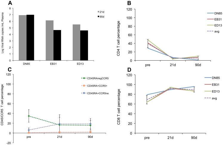
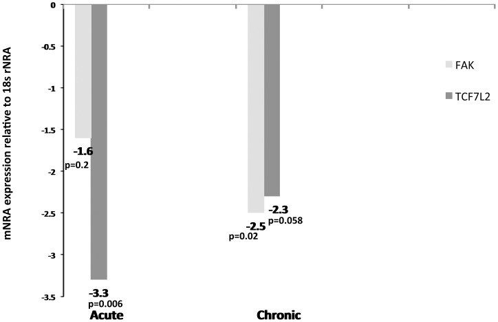
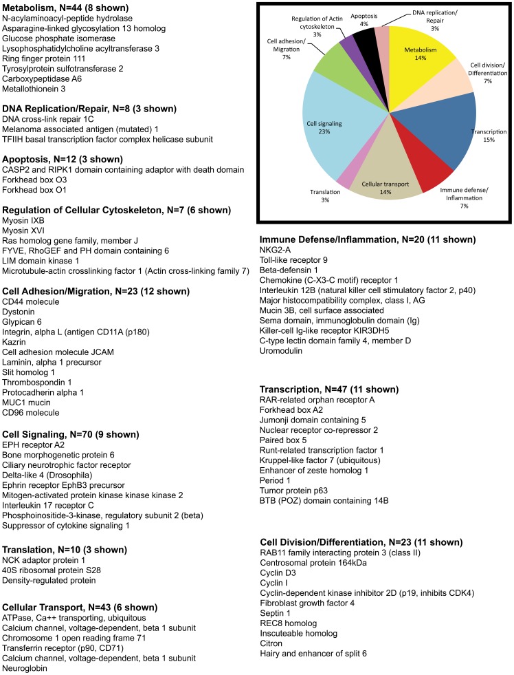
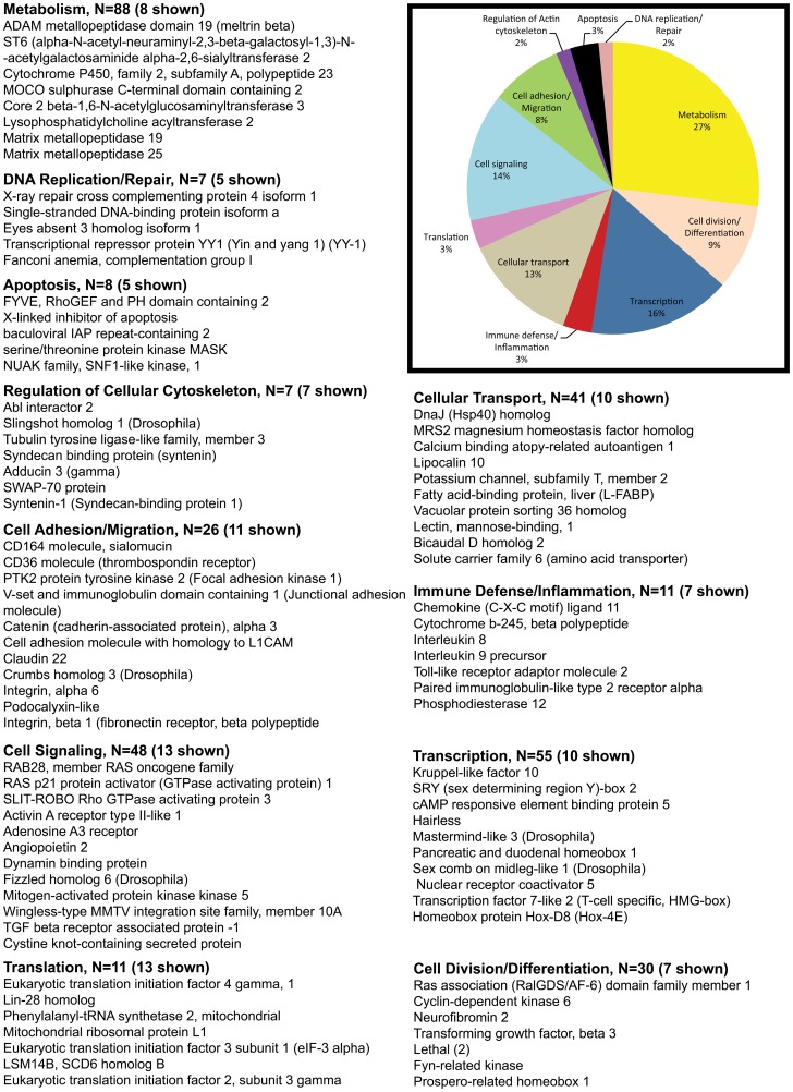
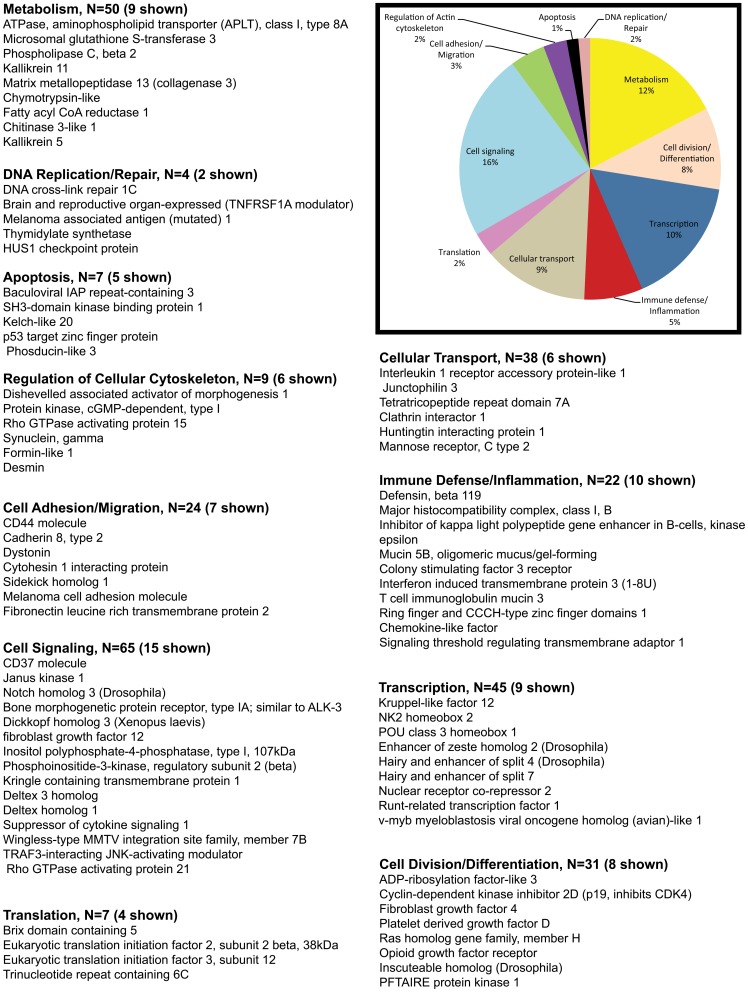
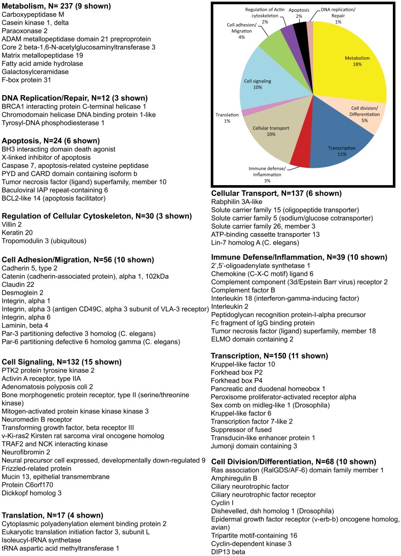
The Gastrointestinal (GI) tract plays a pivotal role in AIDS pathogenesis as it is the primary site for viral transmission, replication and CD4(+) T cell destruction. Accordingly, GI disease (enteropathy) has become a well-known complication and a driver of AIDS progression. To better understand the molecular mechanisms underlying GI disease we analyzed global gene expression profiles sequentially in the intestinal epithelium of the same animals before SIV infection and at 21 and 90 days post infection (DPI). More importantly we obtained sequential excisional intestinal biopsies and examined distinct mucosal components (epithelium. intraepithelial lymphocytes, lamina propria lymphocytes, fibrovascular stroma) separately. Here we report data pertaining to the epithelium. Overall genes associated with epithelial cell renewal/proliferation/differentiation, permeability and adhesion were significantly down regulated (<1.5-7 fold) at 21 and 90DPI. Genes regulating focal adhesions (n = 6), gap junctions (n = 3), ErbB (n = 3) and Wnt signaling (n = 4) were markedly down at 21DPI and the number of genes in each of these groups that were down regulated doubled between 21 and 90DPI. Notable genes included FAK, ITGA6, PDGF, TGFβ3, Ezrin, FZD6, WNT10A, and TCF7L2. In addition, at 90DPI genes regulating ECM-receptor interactions (laminins and ITGB1), epithelial cell gene expression (PDX1, KLF6), polarity/tight junction formation (PARD3B&6B) and histone demethylase (JMJD3) were also down regulated. In contrast, expression of NOTCH3, notch target genes (HES4, HES7) and EZH2 (histone methyltransferase) were significantly increased at 90DPI. The altered expression of genes linked to Wnt signaling together with decreased expression of PDX1, PARD3B, PARD6B and SDK1 suggests marked perturbations in intestinal epithelial function and homeostasis leading to breakdown of the mucosal barrier. More importantly, the divergent expression patterns of EZH2 and JMJD3 suggests that an epigenetic mechanism involving histone modifications may contribute to the massive decrease in gene expression at 90DPI leading to defects in enterocyte maturation and differentiation.
胃肠道(GI)在艾滋病发病机制中起着关键作用,因为它是病毒传播、复制和 CD4(+)T 细胞破坏的主要部位。因此,GI 疾病(肠病)已成为众所周知的并发症和艾滋病进展的驱动因素。为了更好地了解 GI 疾病的分子机制,我们在 SIV 感染前和感染后 21 天和 90 天(DPI)对同一动物的肠道上皮细胞进行了连续的全基因表达谱分析。更重要的是,我们获得了连续的切除性肠道活检,并分别检查了不同的黏膜成分(上皮、上皮内淋巴细胞、固有层淋巴细胞、纤维血管基质)。在这里,我们报告与上皮有关的数据。总体而言,与上皮细胞更新/增殖/分化、通透性和黏附相关的基因在 21 和 90DPI 时显著下调(<1.5-7 倍)。与粘着斑(n=6)、缝隙连接(n=3)、ErbB(n=3)和 Wnt 信号(n=4)相关的基因在 21DPI 时明显下调,这些基因在每个组中的数量在 21 和 90DPI 之间增加了一倍。值得注意的基因包括 FAK、ITGA6、PDGF、TGFβ3、Ezrin、FZD6、WNT10A 和 TCF7L2。此外,在 90DPI 时,调节 ECM-受体相互作用(层粘连蛋白和 ITGB1)、上皮细胞基因表达(PDX1、KLF6)、极性/紧密连接形成(PARD3B 和 6B)和组蛋白去甲基化酶(JMJD3)的基因也下调。相比之下,NOTCH3、NOTCH 靶基因(HES4、HES7)和 EZH2(组蛋白甲基转移酶)的表达在 90DPI 时显著增加。Wnt 信号相关基因表达的改变,以及 PDX1、PARD3B、PARD6B 和 SDK1 的表达降低,表明肠道上皮细胞功能和稳态发生了明显的紊乱,导致黏膜屏障的破坏。更重要的是,EZH2 和 JMJD3 的表达模式的差异表明,涉及组蛋白修饰的表观遗传机制可能导致 90DPI 时大量基因表达的减少,从而导致肠细胞成熟和分化的缺陷。