Cell Cultures Unit, Laboratory of Experimental Surgery and Surgical Research, Faculty of Medicine, Democritus University of Thrace, Dragana, Alexandroupolis, Greece.
Chin Med. 2013 May 4;8(1):9. doi: 10.1186/1749-8546-8-9.
Apigenin (4',5,7-trihydroxyflavone, AP), an active component of many medicinal Chinese herbs, exhibits anticancer properties in vitro and in vivo. This study aims to investigate the genotoxic, cytostatic, and cytotoxic effects of AP and time course changes in the levels of anti- and pro-apoptotic proteins involved in the DNA damage response in HepG2 cells.
The genotoxic potential of AP was determined by sister chromatid exchanges (SCEs) and chromosomal aberrations (CAs) analysis. The levels of cytostaticity and cytotoxicity were evaluated by the proliferation rate and mitotic indices, respectively. MTT was used to study cytotoxicity, while the induction of apoptosis and the expression of apoptosis-related proteins were determined by ELISA.
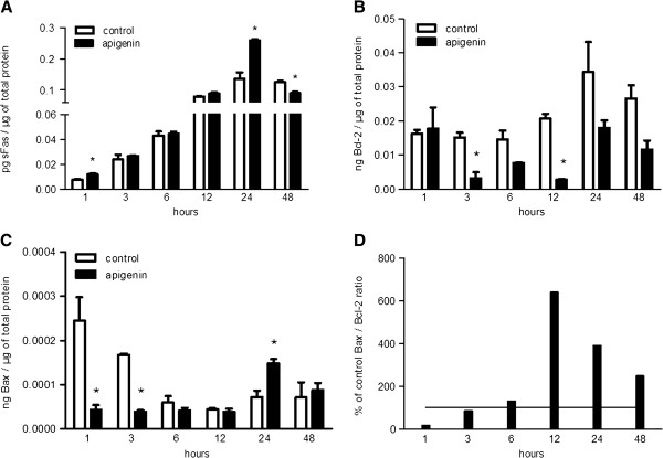
At concentrations greater than 10 μM, AP decreased cell survival in a dose- (48 h: 10 vs. 20 μΜ, P < 0.001 and 20 vs. 50 μΜ, P = 0.005; 72 h: 10 vs. 20 μΜ, P < 0.001 and 20 vs. 50 μΜ, P = 0.001) and time-dependent manner (20 μΜ: 24 vs. 48 h, P < 0.001 and 48 vs. 72 h, P = 0.003; 50 μΜ: 24 vs. 48 h, P < 0.001 and 48 vs. 72 h, P < 0.001; 100 μΜ: 24 vs. 48 h, P < 0.001 and 48 vs. 72 h, P < 0.001). SCEs rates, cell proliferation, and mitotic divisions were also affected in a dose-dependent manner (P < 0.001). There was no change in the frequency of aberrant cells (1 μΜ ΑP: P = 0.554; 10 μM AP: P = 0.337; 20 μΜ AP: P = 0.239). Bcl-2 levels were reduced 3 h after AP administration (P = 0.003) and remained reduced throughout the 48 h observation period (6 h, P = 0.044; 12 h, P = 0.001; 24 h, P = 0.042; 48 h, P = 0.012). Bax and soluble Fas exhibited a transient upregulation 24 h after AP treatment. The Bax/Bcl-2 ratio was also increased at 12 h and remained increased throughout the 48 h observation period.
AP exhibited dose-dependent genotoxic potential in HepG2 cells. The protein levels of sFas, Bcl-2, and Bax were affected by AP to promote cell survival and cell death, respectively.
芹菜素(4',5,7-三羟基黄酮,AP)是许多药用中药的活性成分,在体外和体内均表现出抗癌特性。本研究旨在研究 AP 的遗传毒性、细胞抑制和细胞毒性作用,以及 HepG2 细胞中涉及 DNA 损伤反应的抗凋亡和促凋亡蛋白水平的时程变化。
通过姐妹染色单体交换(SCE)和染色体畸变(CA)分析来确定 AP 的遗传毒性潜力。通过增殖率和有丝分裂指数分别评估细胞抑制和细胞毒性作用。MTT 用于研究细胞毒性,而通过 ELISA 研究细胞凋亡的诱导和凋亡相关蛋白的表达。
AP 浓度大于 10 μM 时,细胞存活率呈剂量依赖性(48 h:10 与 20 μM,P<0.001 和 20 与 50 μM,P=0.005;72 h:10 与 20 μM,P<0.001 和 20 与 50 μM,P=0.001)和时间依赖性(20 μM:24 与 48 h,P<0.001 和 48 与 72 h,P=0.003;50 μM:24 与 48 h,P<0.001 和 48 与 72 h,P<0.001;100 μM:24 与 48 h,P<0.001 和 48 与 72 h,P<0.001)降低。SCE 率、细胞增殖和有丝分裂分裂也呈剂量依赖性(P<0.001)。异常细胞的频率没有变化(1 μM AP:P=0.554;10 μM AP:P=0.337;20 μM AP:P=0.239)。AP 给药后 3 小时 Bcl-2 水平降低(P=0.003),并在整个 48 小时观察期间持续降低(6 小时,P=0.044;12 小时,P=0.001;24 小时,P=0.042;48 小时,P=0.012)。Bax 和可溶性 Fas 显示出 AP 处理后 24 小时的短暂上调。Bax/Bcl-2 比值在 12 小时也增加,并在整个 48 小时观察期间持续增加。
AP 在 HepG2 细胞中表现出剂量依赖性遗传毒性潜力。AP 影响 sFas、Bcl-2 和 Bax 的蛋白水平,分别促进细胞存活和细胞死亡。