Department of Clinical Neurosciences, University of Calgary, Calgary, Canada.
Sensors (Basel). 2013 May 27;13(6):6981-7003. doi: 10.3390/s130606981.
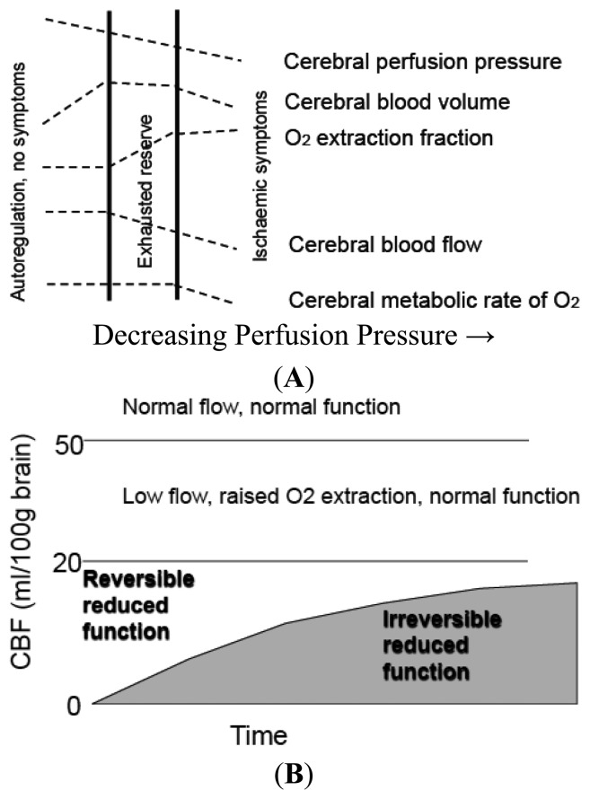
Neuroimaging has improved our understanding of the evolution of stroke at discreet time points helping to identify irreversibly damaged and potentially reversible ischemic brain. Neuroimaging has also contributed considerably to the basic premise of acute stroke therapy which is to salvage some portion of the ischemic region from evolving into infarction, and by doing so, maintaining brain function and improving outcome. The term neurovascular unit (NVU) broadens the concept of the ischemic penumbra by linking the microcirculation with neuronal-glial interactions during ischemia reperfusion. Strategies that attempt to preserve the individual components (endothelium, glia and neurons) of the NVU are unlikely to be helpful if blood flow is not fully restored to the microcirculation. Magnetic resonance imaging (MRI) is the foremost imaging technology able to bridge both basic science and the clinic via non-invasive real time high-resolution anatomical delineation of disease manifestations at the molecular and ionic level. Current MRI based technologies have focused on the mismatch between perfusion-weighted imaging (PWI) and diffusion weighted imaging (DWI) signals to estimate the tissue that could be saved if reperfusion was achieved. Future directions of MRI may focus on the discordance of recanalization and reperfusion, providing complimentary pathophysiological information to current compartmental paradigms of infarct core (DWI) and penumbra (PWI) with imaging information related to cerebral blood flow, BBB permeability, inflammation, and oedema formation in the early acute phase. In this review we outline advances in our understanding of stroke pathophysiology with imaging, transcending animal stroke models to human stroke, and describing the potential translation of MRI to image important interactions relevant to acute stroke at the interface of the neurovascular unit.
神经影像学改善了我们对中风在特定时间点的演变的理解,有助于识别不可逆转损伤和潜在可逆转的缺血性脑。神经影像学也为急性中风治疗的基本前提做出了重要贡献,即从缺血区域中挽救出一部分免于发展为梗死,并通过这种方式,维持脑功能并改善预后。神经血管单元 (NVU) 这一术语通过在缺血再灌注期间将微循环与神经元-胶质相互作用联系起来,拓宽了缺血半影的概念。如果微循环的血流没有完全恢复,试图保护 NVU 的各个组成部分(内皮细胞、胶质细胞和神经元)的策略可能无济于事。磁共振成像 (MRI) 是一种最先进的成像技术,能够通过分子和离子水平的非侵入性实时高分辨率解剖描绘疾病表现,将基础科学和临床联系起来。目前基于 MRI 的技术主要集中在灌注加权成像 (PWI) 和弥散加权成像 (DWI) 信号之间的不匹配上,以估计如果实现再灌注可以挽救的组织。MRI 的未来发展方向可能集中在再通和再灌注之间的不匹配上,为当前梗死核心 (DWI) 和半影 (PWI) 的分区模型提供补充的病理生理学信息,同时提供与脑血流、BBB 通透性、炎症和早期急性阶段水肿形成相关的成像信息。在这篇综述中,我们概述了成像在中风病理生理学理解方面的进展,超越了动物中风模型,涉及人类中风,并描述了 MRI 将成像信息转化为与急性中风在神经血管单元界面相关的重要相互作用的潜力。