Radiology Department, University Medical Center Maribor, Medical Faculty, University of Maribor, Slovenia.
Radiol Oncol. 2013 May 21;47(2):119-24. doi: 10.2478/raon-2013-0009. Print 2013 Jun.
After anterior cruciate ligament (ACL) reconstruction, formation of cortical sclerotic bone encircling the femoral and tibial tunnel is a part of intratunnel graft healing. During the physiological cascades of soft tissue healing and bone growth, cellular and hormonal factors play an important role. The purpose of this study was to non-invasively but quantitatively assess the effect of intraoperatively applied platelet-rich plasma (PRP) on the formation of cortical bone encircling the tibial tunnel.
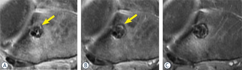
In fifty patients, standard arthroscopic ACL reconstructions were performed. The PRP group (n = 25) received a local application of PRP while the control group (n = 25) did not receive PRP. The proximal tibial tunnel was examined by MRI in the paraxial plane where the portion of the tibial tunnel wall circumference consisting of sclerotic cortical bone was assessed with testing occurring at one, two and a half and six months after surgery.
At one month after surgery, differences between the groups in the amount of cortical sclerotic bone encircling the tunnel were not significant (p = 0.928). At two and a half months, the sclerotic portion of the tunnel wall in the PRP group (36.2%) was significantly larger than in the control (22.5%) group (p = 0.004). At six months, the portion of sclerotic bone in the PRP group (67.1%) was also significantly larger than in the control (53.5%) group (p = 0.003).
Enhanced cortical bone formation encircling the tibial tunnel at 2.5 and 6 months after ACL graft reconstruction results from locally applied platelet-rich plasma.
在前交叉韧带(ACL)重建后,围绕股骨和胫骨隧道的皮质骨硬化形成是隧道内移植物愈合的一部分。在软组织愈合和骨生长的生理级联反应中,细胞和激素因素起着重要作用。本研究的目的是无创但定量评估术中应用富含血小板的血浆(PRP)对胫骨隧道周围皮质骨形成的影响。
在五十名患者中,进行了标准的关节镜 ACL 重建。PRP 组(n = 25)接受 PRP 的局部应用,而对照组(n = 25)未接受 PRP。通过 MRI 在矢状面检查胫骨近端隧道,其中胫骨隧道壁圆周的硬化皮质骨部分用术后 1、2.5 和 6 个月进行的测试进行评估。
术后 1 个月,两组之间围绕隧道的皮质骨硬化量无显著差异(p = 0.928)。术后 2.5 个月,PRP 组隧道壁硬化部分(36.2%)明显大于对照组(22.5%)(p = 0.004)。术后 6 个月,PRP 组硬化骨部分(67.1%)也明显大于对照组(53.5%)(p = 0.003)。
ACL 移植物重建后 2.5 和 6 个月,局部应用富含血小板的血浆可增强胫骨隧道周围皮质骨的形成。